
Abdomen
| Abdomen | |
|---|---|
 | |
 The human abdomen and organs which can be found beneath the surface | |
| Details | |
| Actions | Movement and support for the torso Assistance with breathing Protection for the inner organs Postural support |
| Identifiers | |
| Latin | abdomen |
| Greek | ἦτρον |
| MeSH | D000005 |
| TA98 | A01.1.00.016 |
| TA2 | 127 |
| FMA | 9577 |
| Anatomical terminology | |
The abdomen (colloquially called the belly, tummy, midriff, tucky or stomach) is the part of the body between the
In humans, the abdomen stretches from the thorax at the thoracic diaphragm to the pelvis at the pelvic brim. The pelvic brim stretches from the lumbosacral joint (the intervertebral disc between L5 and S1) to the pubic symphysis and is the edge of the pelvic inlet. The space above this inlet and under the thoracic diaphragm is termed the abdominal cavity. The boundary of the abdominal cavity is the abdominal wall in the front and the peritoneal surface at the rear.
In vertebrates, the abdomen is a large
Contents
The
-
 View of the various organs and blood-vessels in proximity with liver.
 -
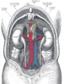 The relations of the viscera and large vessels of the abdomen, seen from behind.


Muscles


(Right) A male abdomen.
There are three layers of muscles in the
The
The
The pyramidalis muscle is small and triangular. It is located in the lower abdomen in front of the rectus abdominis. It originates at the pubic bone and is inserted into the linea alba halfway up to the navel.
Function

Functionally, the human abdomen is where most of the digestive tract is placed and so most of the absorption and digestion of food occurs here. The alimentary tract in the abdomen consists of the lower
.The abdominal wall is split into the posterior (back), lateral (sides), and anterior (front) walls.
Movement, breathing and other functions
The abdominal muscles have different important functions. They assist as
Posture
The transverse abdominis muscle is the deepest muscle; therefore, it cannot be touched from the outside. It can greatly affect the body's posture. The internal obliques are also deep and also affect body posture. Both of them are involved in rotation and lateral flexion of the spine and are used to bend and support the spine from the front. The external obliques are more superficial and are also involved in rotation and lateral flexion of the spine. They also stabilize the spine when upright. The rectus abdominis muscle is not the most superficial abdominal muscle. The tendonous sheath extending from the external obliques cover the rectus abdominis. The rectus abdominis is the muscle that very fit people develop into "six-pack" abs, though there are five vertical sections on each side. The two bottom sections are just above the pubic bone and usually not visible. The rectus abdominals' function is to bend one's back forward (flexion). The main work of the abdominal muscles is to bend the spine forward when contracting concentrically.[4]
Society and culture
Social and cultural perceptions of the outward appearance of the abdomen has varying significance around the world. Depending on the type of society, excess weight can be perceived as an indicator of wealth and prestige due to excess food, or as a sign of poor health due to lack of exercise. In many cultures, bare abdomens are distinctly sexualized and perceived similarly to breast cleavage.
Exercise
.png)
Being key elements of spinal support, and contributors to good posture, it is important to properly exercise the abdominal muscles together with the back muscles because when these are weak or overly tight they can suffer painful spasms and
Clinical significance
Abdominal trauma is an injury to the abdomen and can involve damage to the abdominal organs. There is an associated risk of severe blood loss and infection.[10] Injury to the lower chest can cause injuries to the spleen and liver.[11]
A scaphoid abdomen is when the abdomen is sucked inwards.[12] In a newborn, it may represent a diaphragmatic hernia.[13] In general, it is indicative of malnutrition.[14]
Disease
Many gastrointestinal diseases affect the abdominal organs. These include stomach disease, liver disease, pancreatic disease, gallbladder and bile duct disease; intestinal diseases include enteritis, coeliac disease, diverticulitis, and irritable bowel syndrome.
Examination
Different
Surface landmarks

In the mid-line, a slight furrow extends from the
The upper lateral limit of the abdomen is the subcostal margin (at or near the
One method by which the location of the abdominal contents can be appreciated is to draw three horizontal and two vertical lines.
Horizontal lines

- The highest of the former is the . It corresponds to the first lumbar vertebra behind.
- The second line is the tenth rib). It corresponds to the upper part of the third lumbar vertebra, and it is an inch or so above the umbilicus. It indicates roughly the transverse colon, the lower ends of the kidneys, and the upper limit of the transverse (3rd) part of the duodenum.
- The third line is called the ileo-caecal valve, where the small intestine joins the large intestine.
Vertical lines
The two vertical or mid-Poupart lines are drawn from the point midway between the anterior superior spine and the pubic symphysis on each side, vertically upward to the costal margin.
- The right one is the most valuable, as the ileo-caecal valve is situated where it cuts the intertubercular line. The orifice of the appendix lies an inch lower, at McBurney's point. In its upper part, the vertical line meets the transpyloric line at the lower margin of the ribs, usually the ninth, and here the gallbladderis situated.
- The left mid-Poupart line corresponds in its upper three-quarters to the inner edge of the descending colon.
The right subcostal margin corresponds to the lower limit of the liver, while the right nipple is about half an inch above its upper limit.
Quadrants and regions

The abdomen can be divided into quadrants or regions to describe the location of an organ or structure. Classically, quadrants are described as the left upper, left lower, right upper, and right lower.[citation needed] Quadrants are also often used in describing the site of an abdominal pain.[15]
The abdomen can also be divided into nine regions.
| right hypochondriac/hypochondrium | epigastric/epigastrium | left hypochondriac/hypochondrium |
| right lumbar/flank/ latus /lateral |
umbilical | left lumbar/flank/latus/lateral |
| right inguinal/iliac | pubic |
left inguinal/iliac |
These terms stem from "hypo" meaning "below" and "epi" means "above", while "chondron" means "cartilage" (in this case, the cartilage of the rib) and "gaster" means stomach. The reversal of "left" and "right" is intentional, because the anatomical designations reflect the patient's own right and left.)
The "right iliac fossa" (RIF) is a common site of pain and tenderness in patients who have appendicitis. The fossa is named for the underlying iliac fossa of the hip bone, and thus is somewhat imprecise. Most of the anatomical structures that will produce pain and tenderness in this region are not in fact in the concavity of the ileum. However, the term is in common usage.
Other animals


In
In
The abdomen is sometimes highly modified. In
Unlike other arthropods, insects possess no legs on the abdomen in adult form, though the
In
Abdominal organs can be highly specialized in some animals. For example, the stomach of
See also
- Abdominal fat
References
- ^ Abdomen. Dictionary.com. The American Heritage Dictionary of the English Language, 4th Edition. Retrieved 22 October 2007
- ^ Peritoneum. The Veterinary Dictionary. Elsevier, 2007. Retrieved 22 October 2007
- ^ ISBN 978-1-59339-837-8.)
{{cite encyclopedia}}: CS1 maint: location missing publisher (link - ^ "The Abdominal Muscle Group". Archived from the original on 2015-12-26. Retrieved 2010-07-13.
- ISBN 978-1-4925-3588-1.
- ISBN 978-981-4305-32-7.
- ISBN 978-1-4504-0200-2.
- ISBN 978-1-4504-3416-4.
- ^ Swenson, Doug (2001). "Accumulating strong abs". Power Yoga for Dummies. Hoboken: Wiley Publishers.
- PMID 18436949.
- ISBN 978-0-19-920607-0.
- ISBN 978-1-4160-6257-8.
- ISBN 978-0-415-19184-5.
- ISBN 9780409900774. Retrieved 2013-11-27.
- ISBN 9780071222075.
- ^ "Glossary of Descriptive Terminology". Desertants.org. Archived from the original on 17 May 2013. Retrieved 2013-07-08.
- ^ "Ruminant." The Veterinary Dictionary. Elsevier, 2007. Retrieved 22 October 2007
External links
- . Collier's New Encyclopedia. 1921.
 Media related to Abdomen at Wikimedia Commons