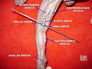
Achilles tendon
| Achilles tendon | |
|---|---|
 The Achilles tendon or calcaneal tendon is attached to the gastrocnemius and soleus muscles. | |
| Details | |
| Location | Back of the lower leg |
| Identifiers | |
| Latin | tendo calcaneus, tendo Achillis |
| MeSH | D000125 |
| TA98 | A04.7.02.048 |
| TA2 | 2662 |
| FMA | 51061 |
| Anatomical terminology | |
The Achilles tendon or heel cord, also known as the calcaneal tendon, is a
Abnormalities of the Achilles tendon include inflammation (Achilles tendinitis), degeneration, rupture, and becoming embedded with cholesterol deposits (xanthomas).
The Achilles tendon was named in 1693 after the Greek hero Achilles.[7]
History
The oldest-known written record of the tendon being named for Achilles is in 1693 by the Flemish/Dutch anatomist Philip Verheyen. In his widely used text Corporis Humani Anatomia he described the tendon's location and said that it was commonly called "the cord of Achilles."[8][9] The tendon has been described as early as the time of Hippocrates, who described it as the "tendo magnus" (Latin for "great tendon")[dubious ] and by subsequent anatomists prior to Verheyen as "chorda Hippocratis" (Latin for "Hippocrates' string").[9]
Verheyen referred to the mythological account of Achilles being held by the heel by his mother
The Achilles tendon is also known as the "tendo calcaneus" (Latin for "calcaneal tendon").
Structure


The Achilles tendon connects muscle to bone, like other
The tendon is covered by the
Along the side of the muscle, and superficial to it, is the small saphenous vein. The sural nerve accompanies the small saphenous vein as it descends in the posterior leg, traveling inferolateral to it as it crosses the lateral border of the Achilles tendon.[12] The tendon is the thickest tendon in the human body.[11] It can receive a load stress 3.9 times body weight during walking and 7.7 times body weight when running.[13]
The blood supply to the Achilles tendon is poor, and mostly via a recurrent branch of the posterior tibial artery, and some through arterial branches passing through surrounding muscles.[11]
Function
Acting via the Achilles tendon, the gastrocnemius and soleus muscles cause
Vibration of the tendon without vision has a major impact on
Clinical significance
Inflammation
It commonly occurs as a result of overuse such as
While
Degeneration
Achilles tendon degeneration (tendinosis) is typically investigated with either
Rupture
Achilles tendon rupture is when the Achilles tendon breaks.[26] Symptoms include the sudden onset of sharp pain in the heel.[18] A snapping sound may be heard as the tendon breaks and walking becomes difficult.[27]
Rupture typically occurs as a result of a sudden bending up of the foot when the calf muscle is engaged, direct
Prevention may include stretching before activity.
Xanthomas
Neurological exam
The Achilles tendon is often tested as part of a
Level or portion of tendon affected:[34]
- Paratendinopathy: The inflammation of a connective tissue sleeve which surrounds the tendon and protects it from friction, irritation, and repeated trauma
- Insertional: Eminently overuse-injury which frequently occurs in running and jumping athletes. Patients affected by insertional Achilles tendinopathy complain of pain on the posterior aspect of the heel and may have morning stiffness, swelling with activity and tenderness at the tendon insertion level.[35] If this condition becomes chronic, calcific deposits at the Achilles insertional level may be developed (due to microfractures and healing of the osteotendinous union) which can degenerate, if it persists over time, in the abnormal bony prominence on the posterior aspect of heel, condition known as Haglund deformity,[36] which can be painful and difficult close-shoes fitting due to friction and irritation.
- Mid-portion: Occurs approximately 2–7 cm proximal from the Achilles insertion[37] into the calcaneus. Characterized by a combination of pain and swelling at this level. It has associated a remarkable impaired performance.
Other animals
The Achilles tendon is short or absent in great apes, but long in arboreal gibbons and humans.[38] It provides elastic energy storage in hopping,[39] walking, and running.[38] Computer models suggest this energy storage Achilles tendon increases top running speed by >80% and reduces running costs by more than three-quarters.[38] It has been suggested that the "absence of a well-developed Achilles tendon in the nonhuman African apes would preclude them from effective running, both at high speeds and over extended distances."[38]
See also
- Heel lifts
References
- S2CID 24159374.
- ^ Richardson TG (1854). Elements of human anatomy: general, descriptive, and practical. Lippincott, Grambo, and Co. pp. 441–.
- ISBN 978-0-683-30471-8.
- ISBN 978-1-55210-001-1.
- ISBN 978-0-7020-5228-6.
- ISBN 978-1-4034-3300-8.
- ISBN 9783319503288.
- ^ Veheyen P (1693), Corporis humani anatomia, Leuven: Aegidium Denique, p. 269, retrieved 12 March 2018,
Vocatum passim chorda Achillis, & ab Hippocrate tendo magnus. (Appendix, caput XII. De musculis pedii et antipedii, p. 269)
- ^ PMID 17463129.
- ISBN 978-1-4160-3165-9.
- ^ ISBN 978-0-8089-2371-8.
- PMID 23099184.
- PMID 10731005.
- ISBN 978-0-8089-2306-0.
- S2CID 12031334.
- S2CID 24715489.
- ^ a b c d e f g Conyer RT, Beahrs T, Kadakia AR (March 2022). "Achilles Tendinitis". OrthoInfo. Retrieved 4 July 2018.
- ^ S2CID 46886582.
- S2CID 3199352.
- PMID 29365222.
- ^ a b c d "Achilles Tendinitis". MSD Manual Professional Edition. March 2018. Retrieved 27 June 2018.
- S2CID 8233009.
- PMID 25981200.
- ^ "Achilles tendinitis — Symptoms and causes". Mayo Clinic. Retrieved 27 June 2018.
- S2CID 8358926.
- ^ ISBN 9780323378222.
- ^ PMID 28613594.
- ^ a b Campagne D (August 2017). "Achilles Tendon Tears". MSD Manual Professional Edition. Retrieved 26 June 2018.
- ^ S2CID 3506883.
- PMID 23659914.
- PMID 18519331.
- ISBN 978-0071748896.
- ISBN 9780729541985.
- ^ Sardon Melo SN (28 August 2018). "Achilles tendinopathy". Emirates Integra. Archived from the original on 4 November 2018. Retrieved 20 October 2020.
- PMID 24559878.
- PMID 24556480.
- S2CID 2214735.
- ^ S2CID 30997461.
- PMID 16326953.
External links
 Media related to Achilles tendon at Wikimedia Commons
