
Amoebiasis
| Amoebiasis | |
|---|---|
| Other names | Amoebic dysentery, amebiasis, entamoebiasis diloxanide furoate, iodoquinoline[2] |
| Frequency | ~480 million[2] |
Amoebiasis, or amoebic dysentery, is an infection of the
Prevention of amoebiasis is by improved
Amoebiasis is present all over the world,
Signs and symptoms
Most infected people, about 90%, are asymptomatic,[9] but this disease has the potential to become serious. It is estimated that about 40,000 to 100,000 people worldwide die annually due to amoebiasis.[4]
Infections can sometimes last for years if there is no treatment. Symptoms take from a few days to a few weeks to develop and manifest themselves, but usually it is about two to four weeks. Symptoms can range from mild diarrhea to dysentery with blood, coupled with intense abdominal pains. Extra-intestinal complications might also arise as a result of invasive infection which includes colitis, liver, lung, or brain abscesses.[9] The blood comes from bleeding lesions created by the amoebae invading the lining of the colon. In about 10% of invasive cases the amoebae enter the bloodstream and may travel to other organs in the body. Most commonly this means the liver,[10] as this is where blood from the intestine reaches first, but they can end up almost anywhere in the body.[citation needed]
Onset time is highly variable and the average asymptomatic infection persists for over a year. It is theorized that the absence of symptoms or their intensity may vary with such factors as strain of amoeba, immune response of the host, and perhaps associated bacteria and viruses.[citation needed]
In asymptomatic infections, the amoeba lives by eating and digesting bacteria and food particles in the gut, a part of the
The ingestion of one viable cyst may cause an infection.[12]
Steroid therapy can occasionally provoke severe amoebic colitis in people with any E. histolytica infection.[13] This bears high mortality: on average more than 50% with severe colitis die.[13]
Cause
Amoebiasis is an infection caused by the
Transmission

Amoebiasis is usually transmitted by the
Amoebic dysentery is one form of
Pathogenesis

Amoebiasis results from tissue destruction induced by the E. histolytica parasite.
E. histolytica causes tissue damage by three main events: direct host cell killing, inflammation, and parasite invasion.[16] The pathogenesis of amoebiasis involves interplay of various molecules secreted by E. histolytica such as LPPG, lectins, cysteine proteases, and amoebapores. Lectins help in the attachment of the parasite to the mucosal layer of the host during invasion. The amoebapores destroy the ingested bacteria present in the colonic environment. Cysteine proteases lyse the host tissues. Other molecules such as PATMK, myosins, G proteins, C2PK, CaBP3, and EhAK1 play an important role in the process of phagocytosis.[17]
Diagnosis
With colonoscopy it is possible to detect small ulcers of between 3–5mm, but diagnosis may be difficult as the mucous membrane between these areas can look either healthy or inflamed.[2] Trophozoites may be identified at the ulcer edge or within the tissue, using immunohistochemical staining with specific anti-E. histolytica antibodies.[7]
Asymptomatic human infections are usually diagnosed by finding cysts shed in the stool. Various flotation or sedimentation procedures have been developed to recover the cysts from fecal matter and stains help to visualize the isolated cysts for microscopic examination. Since cysts are not shed constantly, a minimum of three stools are examined. In symptomatic infections, the motile form (the trophozoite) is often seen in fresh feces. Serological tests exist, and most infected individuals (with symptoms or not) test positive for the presence of antibodies. The levels of antibody are much higher in individuals with liver abscesses. Serology only becomes positive about two weeks after infection. More recent developments include a kit that detects the presence of amoeba proteins in the feces, and another that detects ameba DNA in feces. These tests are not in widespread use due to their expense.[citation needed]
Microscopy is still by far the most widespread method of diagnosis around the world. However it is not as sensitive or accurate in diagnosis as the other tests available. It is important to distinguish the E. histolytica cyst from the cysts of nonpathogenic intestinal protozoa such as
Typically, the organism can no longer be found in the feces once the disease goes extra-intestinal.[citation needed] Serological tests are useful in detecting infection by E. histolytica if the organism goes extra-intestinal and in excluding the organism from the diagnosis of other disorders. An Ova & Parasite (O&P) test or an E. histolytica fecal antigen assay is the proper assay for intestinal infections. Since antibodies may persist for years after clinical cure, a positive serological result may not necessarily indicate an active infection. A negative serological result, however, can be equally important in excluding suspected tissue invasion by E. histolytica.[citation needed]
Stool antigen detection tests have helped to overcome some of the limitations of stool microscopy. Antigen detection tests are easy to use, but they have variable sensitivity and specificity, especially in low-endemic areas.[7]
Polymerase chain reaction (PCR) is considered the gold standard for diagnosis but remains underutilized.[7][18]
-
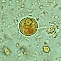 Immature E. histolytica/E. dispar cyst in a concentrated wet mount stained with iodine. This early cyst has only one nucleus and a glycogen mass is visible (brown stain).
 -
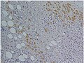
 Amoebae in a colon biopsy from a case of amoebic dysentery.
 -
 Immunohistochemical staining of trophozoites (brown) using specific anti–Entamoeba histolytica macrophage migration inhibitory factor antibodies in a patient with amebic colitis.

.jpg)
_using_specific_anti%E2%80%93Entamoeba_histolytica_macrophage_migration_inhibitory_factor_antibodies_in_a_patient_with_amebic_colitis.jpg)
Prevention

To help prevent the spread of amoebiasis around the home :[citation needed]
- Wash hands thoroughly with soap and hot running water for at least 10 seconds after using the toilet or changing a baby's diaper, and before handling food.
- Clean bathrooms and toilets often; pay particular attention to toilet seats and taps.
- Avoid sharing towels or face washers.
To help prevent infection:[citation needed]
- Avoid raw vegetables when in endemic areas, as they may have been fertilized using human feces.
- Boil water or treat with iodine tablets.
- Avoid eating street foods especially in public places where others are sharing sauces in one container
Good sanitary practice, as well as responsible sewage disposal or treatment, are necessary for the prevention of E. histolytica infection on an endemic level. E.histolytica cysts are usually resistant to chlorination, therefore sedimentation and filtration of water supplies are necessary to reduce the incidence of infection.[9]
E. histolytica cysts may be recovered from contaminated food by methods similar to those used for recovering
Treatment
E. histolytica infections occur in both the intestine and (in people with symptoms) in tissue of the intestine and/or liver.[14] Those with symptoms require treatment with two medications, an amoebicidal tissue-active agent and a luminal cysticidal agent.[9] Individuals that are asymptomatic only need a luminal cysticidal agent.[7]
Prognosis

In the majority of cases, amoebas remain in the gastrointestinal tract of the hosts. Severe ulceration of the gastrointestinal mucosal surfaces occurs in less than 16% of cases. In fewer cases, the parasite invades the soft tissues, most commonly the liver.[10] Only rarely are masses formed (amoebomas) that lead to intestinal obstruction.(Mistaken for Ca caecum and appendicular mass) Other local complications include bloody diarrhea, pericolic and pericaecal abscess.[citation needed]
Complications of hepatic amoebiasis includes subdiaphragmatic abscess, perforation of diaphragm to pericardium and pleural cavity, perforation to abdominal cavital (amoebic peritonitis) and perforation of skin (
Pulmonary amoebiasis can occur from liver lesions by spread through the blood or by perforation of pleural cavity and lung. It can cause lung abscess, pulmono pleural fistula, empyema lung and broncho pleural fistula. It can also reach the brain through blood vessels and cause amoebic brain abscess and amoebic meningoencephalitis. Cutaneous amoebiasis can also occur in skin around sites of colostomy wound, perianal region, region overlying visceral lesion and at the site of drainage of liver abscess.[citation needed]
Urogenital tract amoebiasis derived from intestinal lesion can cause amoebic vulvovaginitis (May's disease), rectovesicle fistula and rectovaginal fistula.[citation needed]
Entamoeba histolytica infection is associated with malnutrition and stunting of growth in children.[20]
Epidemiology
Amoebiasis caused about 55,000 deaths worldwide in 2010, down from 68,000 in 1990.[21][22] In older textbooks it is often stated that 10% of the world's population is infected with Entamoeba histolytica.[citation needed] Nevertheless, this means that there are up to 50 million true E. histolytica infections and approximately seventy thousand die each year, mostly from liver abscesses or other complications. Although usually considered a tropical parasite, the first case reported (in 1875) was actually in St Petersburg in Russia, near the Arctic Circle.[23] Infection is more common in warmer areas, but this is because of both poorer hygiene and the parasitic cysts surviving longer in warm moist conditions.[14]
History
Amoebiasis was first described by Fedor A. Lösch in 1875, in northern Russia.
The Nicobarese people have attested to the medicinal properties found in Glochidion calocarpum, a plant common to India, saying that its bark and seed are most effective in curing abdominal disorders associated with amoebiasis.[27]
Society and culture
An outbreak of amoebic dysentery occurs in Diana Gabaldon's novel A Breath of Snow and Ashes.[28]
References
- ^ "Entamoebiasis". Medical Subject Headings. National Library of Medicine. Retrieved 9 April 2024.
- ^ ISBN 978-0-7020-5101-2.
- ^ a b c "General Information | Amebiasis | Parasites | CDC". www.cdc.gov. 29 December 2021. Retrieved 13 September 2022.
- ^ S2CID 203849436.
- S2CID 248741177.
- ISBN 978-1-118-73456-8.
- ^ PMID 30046644.
- ISBN 978-1-119-60016-9.
- ^ S2CID 218475533.
- ^ PMID 26088504.
- ISBN 978-0-632-04204-3.
- Bad Bug Book. United States Food and Drug Administration: Center for Food Safety & Applied Nutrition. 2007-12-28. Archivedfrom the original on 9 July 2009. Retrieved 2009-07-13.
- ^ PMID 27467600.
- ^ ISBN 978-0-8385-8529-0.
- ^ "safewateronline.com: Travelers' Diarrhea". Archived from the original on 6 June 2008. Retrieved 2020-06-28.
- PMID 31008019.
- PMID 6085216.
- PMID 31674299.
- ^ "FDA Bacteriological Analytical Manual". Archived from the original on 2008-04-06. Retrieved 2008-03-26.
- PMID 16730764.
- PMID 31725715.
- S2CID 1541253.
- .
- PMID 3524005.
- ^ "Water and Waste Systems". Archived from the original on 2017-01-19. Retrieved 2017-01-19.[unreliable medical source?]
- PMID 12631887.
- S2CID 32113455.
- ^ "Outlandish Observations". Retrieved 2020-11-24.[unreliable medical source?]
External links
| Authority control databases: National |
|---|