
Amyloidosis
| Amyloidosis | |
|---|---|
| Prognosis | Improved with treatment[3] |
| Frequency | 3–13 per million per year (AL amyloidosis)[2] |
| Deaths | 1 per 1,000 people (developed world)[3] |
Amyloidosis is a group of diseases in which abnormal
There are about 36 different types of amyloidosis, each due to a specific
Diagnosis may be suspected when
Treatment is geared towards decreasing the amount of the involved protein.
Signs and symptoms

The presentation of amyloidosis is broad and depends on the site of amyloid accumulation. The kidney and heart are the most common organs involved.
Kidneys
Heart
Amyloid deposition in the heart can cause both diastolic and systolic
Nervous system
People with amyloidosis may have central nervous system involvement,[13] along with peripheral involvement which causes sensory and autonomic neuropathies. Sensory neuropathy develops in a symmetrical pattern and progresses in a distal to proximal manner. Autonomic neuropathy can present as orthostatic hypotension but may manifest more gradually with nonspecific gastrointestinal symptoms like constipation, nausea, or early satiety.[10] Amyloidosis of the central nervous system can have more severe and systemic presentations that may include life-threatening arrhythmias, cardiac failure, malnutrition, infection, or death.[14]
Neuropathic presentation can depend on the etiology of amyloidosis.[14] People with amyloidosis may experience dysfunction in various organ systems depending on the location and extent of nervous system involvement.[8] For example, peripheral neuropathy can cause erectile dysfunction, incontinence and constipation, pupillary dysfunction, and sensory loss depending on the distribution of amyloidosis along different peripheral nerves.[14]
Gastrointestinal and accessory organs
Accumulation of amyloid proteins in the gastrointestinal system may be caused by a wide range of amyloid disorders and have different presentations depending on the degree of organ involvement.[15] Potential symptoms include weight loss, diarrhea, abdominal pain, heartburn (gastrointestinal reflux), and GI bleeding.[15] Amyloidosis may also affect accessory digestive organs including the liver, and may present with jaundice, fatty stool, anorexia, fluid buildup in the abdomen, and spleen enlargement.[15]
Accumulation of amyloid proteins in the liver can lead to elevations in serum
Glands
Both the
"Amyloid deposits occur in the
Musculoskeletal system
Amyloid proteins deposit most commonly inside the knee, followed by hands, wrists, elbow, hip, and ankle, causing joint pain.[16] In males with advanced age (>80 years), there is significant risk of wild-type transthyretin amyloid deposition in synovial tissue of knee joint, but predominantly in old age deposition of wild type transthyretin is seen in cardiac ventricles. ATTR deposits have been found in ligamentum flavum of patients that underwent surgery for lumbar spinal stenosis.[17]
In beta 2-microglobulin amyloidosis, males have high risk of getting carpal tunnel syndrome.[18] Aβ2MG amyloidosis (Hemodialysis associated amyloidosis) tends to deposit in synovial tissue, causing chronic inflammation of the synovial tissue in knee, hip, shoulder and interphalangeal joints.[18] Amyloid light chains deposition in shoulder joint causes enlarged shoulders, also known as "shoulder pad sign".[18] Amyloid light chain depositions can also cause bilateral symmetric polyarthritis.[18]
The deposition of amyloid proteins in the bone marrow without causing
Eyes
A rare development is amyloid purpura, a susceptibility to bleeding with bruising around the eyes, termed "raccoon-eyes". Amyloid purpura is caused by amyloid deposition in the blood vessels and reduced activity of thrombin and factor X, two clotting proteins that lose their function after binding with amyloid.[10]
Oral cavity
Amyloid deposits in tissue can cause enlargement of structures. Twenty percent of people with AL amyloidosis have an enlarged tongue, that can lead to obstructive sleep apnea, difficulty swallowing, and altered taste.[11] Tongue enlargement does not occur in ATTR or AA amyloidosis.[10] Deposition of amyloid in the throat can cause hoarseness.[10]
Pathogenesis
Amyloidoses can be considered
Amyloid-forming proteins aggregate into distinctive fibrillar forms with a
Diagnosis
Diagnosis of amyloidosis generally requires tissue biopsy.[2] The biopsy is assessed for evidence of characteristic amyloid deposits. The tissue is treated with various stains. The most useful stain in the diagnosis of amyloid is Congo red, which, combined with polarized light, makes the amyloid proteins appear apple-green on microscopy. Also, thioflavin T stain may be used.[22] A number of imaging techniques such as a Nuclear Medicine PYP scan, DPD scan or SAP scan are also in use.[23]
A sample of tissue can be biopsied or obtained directly from the affected internal organ, but the first-line site of biopsy is subcutaneous abdominal fat, known as a "fat pad biopsy", due to its ease of acquisition.[24][25] An abdominal fat biopsy is not completely sensitive and may result in false negatives, which means a negative result does not exclude the diagnosis of amyloidosis.[24][25] However, direct biopsy of the affected organ may still be unnecessary as other less invasive methods of biopsy can also be used, including rectal mucosa, salivary gland, lip, or bone marrow biopsy which can achieve a diagnosis in up to 85% of people.[24]
In the amyloid deposition of the joints, there will be a decreased signal in both
The type of the amyloid protein can be determined in various ways: the detection of abnormal proteins in the bloodstream (on
AL was previously considered the most common form of amyloidosis, and a diagnosis often begins with a search for
ATTR is now considered to be the most common form of amyloidosis. It may be either age related in wild-type ATTR (ATTRv) or familial transthyretin-associated amyloidosis, is suspected in people with family history of idiopathic neuropathies or heart failure who lack evidence of plasma cell dyscrasias. ATTR can be identified using isoelectric focusing which separates mutated forms of transthyretin. Findings can be corroborated by genetic testing to look for specific known mutations in transthyretin that predispose to amyloidosis.[10]
AA is suspected on clinical grounds in individuals with longstanding infections or inflammatory diseases. AA can be identified by immunohistochemistry staining.[10]
-
 Small bowel duodenum with amyloid deposition Congo red 10X
 -
 Amyloidosis, dystrophic calcification
 -
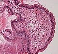 Small bowel duodenum with amyloid deposition 20X
 -
 Amyloidosis, Node, Congo Red
 -
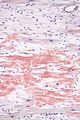
 Amyloidosis, blood vessels, H&E
 -
 Amyloidosis, lymph node, H&E
 -
 Amyloidosis, lymph node, polarizer
 -

-
 Congo red stain.


Classification
Historical classification systems were based on clinical factors. Until the early 1970s, the idea of a single amyloid substance predominated. Various descriptive classification systems were proposed based on the organ distribution of amyloid deposits and clinical findings. Most classification systems included primary (i.e.,
The modern era of amyloidosis classification began in the late 1960s with the development of methods to make amyloid fibrils soluble. These methods permitted scientists to study the chemical properties of amyloids.[medical citation needed] Descriptive terms such as primary amyloidosis, secondary amyloidosis, and others (e.g., senile amyloidosis), which are not based on cause, provide little useful information and are no longer recommended.
The modern classification of amyloid disease tends to use an abbreviation of the protein that makes the majority of deposits, prefixed with the letter A. For example, amyloidosis caused by
Other forms are due to different diseases causing overabundant or abnormal protein production – such as with overproduction of
About 60 amyloid proteins have been identified so far.[27] Of those, at least 36 have been associated with a human disease.[28]
All amyloid fibril proteins start with the letter "A" followed by the protein suffix (and any applicable specification). See below for a list of amyloid fibril proteins which have been found in humans:[29]
| Fibril protein | Precursor protein | Target Organs | Systemic and/or localized | Acquired or hereditary |
|---|---|---|---|---|
| AL | Immunoglobulin light chain | All organs, usually except CNS | S, L | A, H |
| AH | Immunoglobulin heavy chain | All organs except CNS | S, L | A |
| AA | (Apo) serum amyloid A | All organs except CNS | S | A |
| ATTR | Transthyretin, wild type
Transthyretin, variants |
Heart mainly in males, lung, ligaments, tenosynovium
PNS, ANS, heart, eye, leptomeninges |
S
S |
A
H |
| Aβ2M | β2-microglobulin, wild type
β2-microglobulin, variants |
Musculoskeletal system
ANS |
S
S |
A
H |
| AApoAI | Apolipoprotein A I , variants
|
Heart, liver, kidney, PNS, testis, larynx (C
terminal variants), skin (C terminal variants) |
S | H |
| AApoAII | Apolipoprotein A II , variants
|
Kidney | S | H |
| AApoAIV | Apolipoprotein A IV, wild type | Kidney medulla and systemic | S | A |
| AApoCII | Apolipoprotein C II, variants | Kidney | S | H |
| AApoCIII | Apolipoprotein C III, variants | Kidney | S | H |
| AGel | Gelsolin, variants | Kidney, PNS, cornea | S | H |
| ALys | Lysozyme, variants | Kidney | S | H |
| ALECT2 | Leukocyte chemotactic factor-2 | Kidney, primarily | S | A |
| AFib | Fibrinogen a, variants | Kidney, primarily | S | H |
| ACys | Cystatin C, variants | CNS, PNS, skin | S | H |
| ABri | ABriPP, variants | CNS | S | H |
| ADanb | ADanPP, variants | CNS | L | H |
| Aβ | Aβ protein precursor, wild type
Aβ protein precursor, variant |
CNS | L
L |
A
H |
| AαSyn | α-Synuclein | CNS | L | A |
| ATau | Tau | CNS | L | A |
| APrP | Prion protein, wild type
Prion protein variants Prion protein variant |
CJD, fatal insomnia
CJD, GSS syndrome, fatal insomnia PNS |
L
L S |
A
H H |
| ACal | (Pro)calcitonin | C-cell thyroid tumours
Kidney |
L
S |
A
A |
| AIAPP | Islet amyloid polypeptidec | Islets of Langerhans, insulinomas | L | A |
| AANF | Atrial natriuretic factor | Cardiac atria | L | A |
| APro | Prolactin | Pituitary prolactinomas, aging pituitary | L | A |
| AIns | Insulin | Iatrogenic, local injection | L | A |
| ASPCd | Lung surfactant protein | Lung | L | A |
| ACor | Corneodesmosin | Cornified epithelia, hair follicles | L | A |
| AMed | Lactadherin | Senile aortic, media | L | A |
| AKer | Kerato-epithelin | Cornea, hereditary | L | A |
| ALac | Lactoferrin | Cornea | L | A |
| AOAAP | Odontogenic ameloblast-associated protein | Odontogenic tumours | L | A |
| ASem1 | Semenogelin 1 | Vesicula seminalis | L | A |
| AEnf | Enfurvitide | Iatrogenic | L | A |
| ACatKe | Cathepsin K | Tumour associated | L | A |
| AEFEMP1e | EGF-containing fibulin-like extracellular
matrix protein 1 (EFEMP1) |
Portal veins, Aging associated | L | A |
Alternative
An older clinical method of classification refers to amyloidoses as systemic or localised:
- Systemic amyloidoses affect more than one body organ or system. Examples are AL, AA and Aβ2m.[30]
- Localised amyloidoses affect only one body organ or tissue type. Examples are medullary carcinoma of the thyroid)[30]
Another classification is primary or secondary.[medical citation needed]
- Primary amyloidoses arise from a disease with disordered immune cell function, such as multiple myelomaor other immunocyte dyscrasias.
- Secondary (reactive) amyloidoses occur as a complication of some other chronic inflammatory or tissue-destroying disease. Examples are reactive systemic amyloidosis and secondary cutaneous amyloidosis.[30]
Additionally, based on the tissues in which it is deposited, it is divided into mesenchymal (organs derived from mesoderm) or parenchymal (organs derived from ectoderm or endoderm).[medical citation needed]
Treatment
Treatment depends on the type of amyloidosis that is present. Treatment with high dose melphalan, a chemotherapy agent, followed by stem cell transplantation has shown promise in early studies and is recommended for stage I and II AL amyloidosis.[26] However, only 20–25% of people are eligible for stem cell transplant. Chemotherapy treatment including cyclophosphamide-bortezomib-dexamethasone is currently the recommended treatment option for people with AL Amyloidosis not eligible for transplant.[5]
In AA, symptoms may improve if the underlying condition is treated. In people who have inflammation caused by AA amyloidosis, tumour necrosis factor (TNF)-alpha inhibitors such as infliximab and etanercept are used for an average duration of 20 months. If TNF-alpha inhibitors are not effective, Interleukin-1 inhibitors (e.g., anakinra, canakinumab, rilonacept) and interleukin-6 inhibitors (e.g., tocilizumab) may be considered.[31]
Management of ATTR amyloidosis will depend on its classification as wild type or variant.[5] Both may be treated with tafamidis, a low toxicity oral agent that prevents destabilization of correctly folded protein.[5] Studies showed tafamidis reduced mortality and hospitalization due to heart failure.[5] Previously, for variant ATTR amyloidosis, liver transplant was the only effective treatment.[5] New therapies include diflunisal, inotersen, and patisiran.
Diflunisal binds to misfolded mutant TTR protein to prevent its buildup, like how tafamidis works. Low-certainty evidence indicates that it mitigates worsening of peripheral neuropathy and disability from disease progression.[32]
Inotersen blocks gene expression of both wild-type and mutant TTR, reducing amyloid precursor. Moderate-certainty evidence suggests that it mitigates worsening of peripheral neuropathy. Long-term efficacy and safety of inotersen use in people with mutant TTR-related amyloidosis is still be evaluated in a phase-III clinical trial as of 2021. Both diflunisal and inotersen may also mitigate declines in quality-of-life, though the evidence for this effect is unclear.[32] For people with cardiac ATTR the effect of inotersen use is inconclusive and requires further investigation.[33] In 2018, inotersen was approved by the European Medicines Agency to treat polyneuropathy in adults with hereditary transthyretin amyloidosis.[34] It has since been approved for use in Canada, the European Union and in the USA.[35]
Patisiran functions similarly to inotersen. Moderate-certainty evidence suggests that patisiran mitigates worsening of peripheral neuropathy and disability from disease progression. Additionally, low-certainty evidence suggests that patisiran mitigates decreases in quality-of-life and slightly reduces the rate of adverse events versus placebo. There is no evidence of an effect on mortality rate.[32] A review of early data from use of patisiran in people with variant cardiac ATTR suggests that it may reduce mortality and hospitalization, however this is still being investigated and requires further investigation.[33] In 2018, patisiran was not recommended by NICE in the UK for hereditary transthyretin-related amyloidosis.[36] As of July 2019 further review however is occurring.[37] It was approved for this use in the United States, however.[38]
The roles of inotersen and patisiran in cardiac ATTR amyloidosis are still being investigated.[5]
In 2021, in a clinical trial using the CRISPR gene-editing technique, several participants had an "80% to 96% drop in TTR levels, on par or better than the average of 81%" who were given patisiran.[39]
Vutrisiran was approved by the U.S. Food and Drug Administration (FDA) in June 2022, for the treatment of the polyneuropathy of hereditary transthyretin-mediated (hATTR) amyloidosis in adults.[40]
Support groups
People affected by amyloidosis are supported by organizations, including the Amyloidosis Research Consortium, Amyloidosis Foundation, Amyloidosis Support Groups, and Australian Amyloidosis Network.[41][42]
Prognosis
Prognosis varies with the type of amyloidosis and the affected organ system. Prognosis for untreated AL cardiac amyloidosis is poor, with a median survival of six months.[43] More specifically, AL amyloidosis can be classified as stage I, II or III based on cardiac biomarkers like Nt-proBNP and cardiac troponin.[44] Survival diminishes with increasing stage, but recent advancements in treatments have improved median survival rates for stages I, II, and III, to 91.2, 60, and 7 months respectively.[44]
Outcomes in a person with AA amyloidosis depend on the underlying disease, organ(s) affected, and correlate with the concentration of serum amyloid A protein.[5]
People with ATTR, mutant ATTR and wild-type ATTR have a better prognosis when compared to people with AL and may survive for over a decade.[10][45] Survival time is not associated with gender or age, however, some measures of reduced heart function are associated with a shorter survival time.[45]
Senile systemic amyloidosis was determined to be the primary cause of death for 70% of people over 110 who have been autopsied.[46][47]
Epidemiology
Amyloidosis has a combined estimated prevalence of 30 per 100,000 persons with the three most common forms being AL, ATTR, and AA.[48] The median age at diagnosis is 64.[11]
AL has the highest incidence at approximately 12 cases per million persons per year and an estimated prevalence of 30,000 to 45,000 cases in the US and European Union.[48][5]
AA amyloidoses is the most common form in developing countries and can complicate longstanding infections with tuberculosis, osteomyelitis, and bronchiectasis. AA amyloidosis is caused by an increase in extracellular deposition of serum amyloid A (SAA) protein. SAA protein levels can rise in both direct and indirect manners, through infection, inflammation, and malignancies.[49] The most common causes of AA amyloidosis in the West are rheumatoid arthritis, inflammatory bowel disease, psoriasis, and familial Mediterranean fever.[10]
People undergoing long-term hemodialysis (14–15 years) can develop amyloidosis from accumulation of light chains of the HLA 1 complex which is normally filtered out by the kidneys.[11]
Wild-type transthyretin (ATTR) amyloidosis is found in a quarter of elderly at postmortem.[50] ATTR is found in 13–19% of people experiencing heart failure with preserved ejection fraction, making it a very common form of systemic amyloidosis.[51]
Research
Treatments for ATTR-related
See also
References
- ^ Hawkins P (29 April 2015). "AL amyloidosis". Wikilite.com. Archived from the original on 22 December 2015. Retrieved 19 December 2015.
- ^ S2CID 215069282.
- ^ PMID 16409147.
- ^ "AL amyloidosis". rarediseases.info.nih.gov. Genetic and Rare Diseases Information Center (GARD). Archived from the original on 24 April 2017. Retrieved 22 April 2017.
- ^ S2CID 220385853.
- ^ S2CID 218600304.
- PMID 23425518.
- ^ a b c "Amyloidosis & Kidney Disease". National Institute of Diabetes and Digestive and Kidney Diseases. U.S. Department of Health and Human Services. Archived from the original on 19 November 2021. Retrieved 19 November 2021.
- ^ a b Lewis JB, Neilson EG (2018). "Glomerular Diseases". In Jameson J, Fauci AS, Kasper DL, Hauser SL, Longo DL, Loscalzo J (eds.). Harrison's Principles of Internal Medicine (20 ed.). McGraw Hill. Archived from the original on 29 November 2021. Retrieved 29 November 2021.
- ^ PMID 9302305.
- ^ S2CID 25431033.
- ^ PMID 30509186.
- ^ Soprano DR, Herbert J, Soprano KJ, Schon EA, Goodman DS. Demonstration of transthyretin mRNA in the brain and other extrahepatic tissues in the rat. J Biol Chem 1985; 260 (21) 11793-11798
- ^ S2CID 204850185.
- ^ PMID 28611935.
- ^ PMID 27153791.
- PMID 33274477.
- ^ PMID 29762107.
- ^ PMID 28498720.
- ^ PMID 21483018.
- OCLC 654382006.
- ^ from the original on 5 December 2011.
- PMID 23896486.
- ^ from the original on 14 June 2022. Retrieved 25 December 2020.
- ^ S2CID 4762107.
- ^ PMID 22100031.
- PMID 17223074.
- S2CID 31198109.
- S2CID 225073269.
- ^ ISBN 978-1-4160-2973-1. 8th edition.
- S2CID 18876892.
- ^ PMID 32311072.
- ^ PMID 32969287.
- ^ "Tegsedi". Europeans Medicines Agency. Archived from the original on 8 October 2020. Retrieved 12 March 2021.
- PMID 31118583.
- ^ "Patisiran for treating hereditary transthyretinrelated amyloidosis". Archived from the original on 4 July 2019. Retrieved 20 July 2019.
- ^ "Patisiran for treating hereditary transthyretin-related amyloidosis [ID1279] | Guidance | NICE". National Institute for Health and Care Excellence (Nice). Archived from the original on 20 July 2019. Retrieved 20 July 2019.
- S2CID 52813638.
- S2CID 235722446.
- ^ "Alnylam Announces FDA Approval of Amvuttra (vutrisiran), an RNAi Therapeutic for the Treatment of the Polyneuropathy of Hereditary Transthyretin-Mediated Amyloidosis in Adults". Alnylam. 13 June 2022. Archived from the original on 14 June 2022. Retrieved 14 June 2022 – via Business Wire.
- ^ "Amyloidosis - NORD (National Organization for Rare Disorders)". NORD (National Organization for Rare Disorders). Archived from the original on 16 March 2016. Retrieved 15 March 2016.
- ^ "Amyloidosis primary cutaneous – Disease – Organizations – Genetic and Rare Diseases Information Center (GARD) – NCATS Program". rarediseases.info.nih.gov. Archived from the original on 15 March 2016. Retrieved 15 March 2016.
- PMID 29222231.
- ^ PMID 27634125.
- ^ S2CID 73419672.
- PMID 22579241.
- ^ "Searching for the Secrets of the Super Old". Science. 26 September 2008. pp. 1764–1765. Archived from the original on 9 March 2013. Retrieved 22 February 2013.
- ^ S2CID 205541963.
- S2CID 208299494.
- from the original on 14 June 2022. Retrieved 18 March 2022.
- PMID 33993188.
- S2CID 49658028.
- ^ Office of the Commissioner. "Press Announcements - FDA approves first-of-its kind targeted RNA-based therapy to treat a rare disease". www.fda.gov. Archived from the original on 7 September 2018. Retrieved 11 August 2018.
- ^ PMID 32587078.
External links
- Amyloidosis at Curlie