
Anterior cruciate ligament
| Anterior cruciate ligament | |
|---|---|
Intercondyloid eminence of the tibia | |
| Identifiers | |
| Latin | ligamentum cruciatum anterius |
| MeSH | D016118 |
| TA98 | A03.6.08.007 |
| TA2 | 1890 |
| FMA | 44614 |
| Anatomical terminology] | |
The anterior cruciate ligament (ACL) is one of a pair of
Structure
The ACL originates from deep within the notch of the
Purpose
The purpose of the ACL is to resist the motions of anterior tibial translation and internal tibial rotation; this is important to have rotational stability.
Clinical significance
Injury

An ACL tear is one of the most common knee injuries, with over 100,000 tears occurring annually in the US.
Nonoperative treatment of the ACL
ACL reconstruction is the most common treatment for an ACL tear, but it is not the only treatment available for individuals. Some may find it more beneficial to complete a nonoperative rehabilitation program. Individuals who are going to continue with physical activity that involves cutting and pivoting, and individuals who are no longer participating in those specific activities both are candidates for the nonoperative route.[13] In comparing operative and nonoperative approaches to ACL tears, few differences were noted between surgical and nonsurgical groups, with no significant differences in regard to knee function or muscle strength reported by the patients.[citation needed]
The main goals to achieve during rehabilitation (rehab) of an ACL tear is to regain sufficient functional stability, maximize full muscle strength, and decrease risk of reinjury.[citation needed] Typically, three phases are involved in nonoperative treatment - the acute phase, the neuromuscular training phase, and the return to sport phase. During the acute phase, the rehab is focusing on the acute symptoms that occur right after the injury and are causing an impairment. The use of therapeutic exercises and appropriate therapeutic modalities is crucial during this phase to assist in repairing the impairments from the injury. The neuromuscular training phase is used to focus on the patient regaining full strength in both the lower extremity and the core muscles. This phase begins when the patient regains full range of motion, no effusion, and adequate lower extremity strength. During this phase, the patient completes advanced balance, proprioception, cardiovascular conditioning, and neuromuscular interventions.[13] In the final, return to sport phase, the patient focuses on sport-specific activities and agility. A functional performance brace is suggested to be used during the phase to assist with stability during pivoting and cutting activities.[13]
Operative treatment of the ACL
Anterior cruciate ligament surgery is a complex operation that requires expertise in the field of orthopedic and sports medicine. Many factors should be considered when discussing surgery, including the athlete's level of competition, age, previous knee injury, other injuries sustained, leg alignment, and graft choice. Typically, four graft types are possible, the bone-patella tendon-bone graft, the semitendinosus and gracilis tendons (quadrupled hamstring tendon), quadriceps tendon, and an allograft.[14] Although extensive research has been conducted on which grafts are the best, the surgeon typically chooses the type of graft with which he or she is most comfortable. If rehabilitated correctly, the reconstruction should last. In fact, 92.9% of patients are happy with graft choice.[14]
Prehabilitation has become an integral part of the ACL reconstruction process. This means that the patient exercises before getting surgery to maintain factors such as range of motion and strength. Based on a single leg hop test and self-reported assessment, prehab improved function; these effects were sustained 12 weeks postoperatively.[15]
Postsurgical rehabilitation is essential in the recovery from the reconstruction. This typically takes a patient 6 to 12 months to return to life as it was prior to the injury.[16] The rehab can be divided into protection of the graft, improving range of motion, decrease swelling, and regaining muscle control.[16] Each phase has different exercises based on the patients' needs. For example, while the ligament is healing, a patient's joint should not be used for full weight-bearing, but the patient should strengthen the quadriceps and hamstrings by doing quad sets and weight shifting drills. Phase two would require full weight-bearing and correcting gait patterns, so exercises such as core strengthening and balance exercises would be appropriate. In phase three, the patient begins running, and can do aquatic workouts to help with reducing joint stresses and cardiorespiratory endurance. Phase four includes multiplanar movements, thus enhancing a running program and beginning agility and plyometric drills. Lastly, phase five focuses on sport- or life-specific motions, depending on the patient.[16]
A 2010 Los Angeles Times review of two medical studies discussed whether ACL reconstruction was advisable. One study found that children under 14 who had ACL reconstruction fared better after early surgery than those who underwent a delayed surgery. For adults 18 to 35, though, patients who underwent early surgery followed by rehabilitation fared no better than those who had rehabilitative therapy and a later surgery.[17]
The first report focused on children and the timing of an ACL reconstruction. ACL injuries in children are a challenge because children have open growth plates in the bottom of the femur or thigh bone and on the top of the tibia or shin. An ACL reconstruction typically crosses the growth plates, posing a theoretical risk of injury to the growth plate, stunting leg growth, or causing the leg to grow at an unusual angle.[18]
The second study noted focused on adults. It found no significant statistical difference in performance and pain outcomes for patients who receive early ACL reconstruction vs. those who receive physical therapy with an option for later surgery. This would suggest that many patients without instability, buckling, or giving way after a course of rehabilitation can be managed nonoperatively, but was limited to outcomes after two years and did not involve patients who were serious athletes.[17] Patients involved in sports requiring significant cutting, pivoting, twisting, or rapid acceleration or deceleration may not be able to participate in these activities without ACL reconstruction.[19]
ACL injuries in women
Risk differences between outcomes in men and women can be attributed to a combination of multiple factors, including anatomical, hormonal, genetic, positional, neuromuscular, and environmental factors.[20] The size of the anterior cruciate ligament is often the most reported difference. Studies look at the length, cross-sectional area, and volume of ACLs. Researchers use cadavers, and in vivo placement to study these factors, and most studies confirm that women have smaller anterior cruciate ligaments. Other factors that could contribute to higher risks of ACL tears in women include patient weight and height, the size and depth of the intercondylar notch, the diameter of the ACL, the magnitude of the tibial slope, the volume of the tibial spines, the convexity of the lateral tibiofemoral articular surfaces, and the concavity of the medial tibial plateau.[21] While anatomical factors are most talked about, extrinsic factors, including dynamic movement patterns, might be the most important risk factor when it comes to ACL injury.[22]
Gallery
-
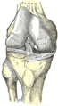 Right knee joint, from the front, showing interior ligaments
 -
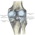
 Left knee joint from behind, showing interior ligaments
 -
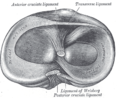 Head of right tibia seen from above, showing menisci and attachments of ligaments
 -
 Capsule of right knee-joint (distended), posterior aspect
 -
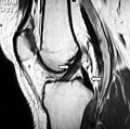 MRI shows normal signal of both cruciate ligaments (arrows)
 -
 Knee joint, deep dissection, anteromedial view


See also
References
- ^ "Canine Cranial Cruciate Ligament Disease" (PDF). Melbourne Veterinary Referral Centre. pp. 1–2. Archived from the original (PDF) on 19 July 2008. Retrieved September 8, 2009.
- PMID 3969275.
- S2CID 45919449.
- S2CID 25658911.
- PMID 17075382.
- ^ PMID 23015856.
- ^ a b c Liu-Ambrose, T. (December 2003). "The anterior cruciate ligament and functional stability of the knee joint". British Columbia Medical Journal. 45 (10): 495–499. Retrieved 2018-11-15.
- PMID 20949884.
- ^ a b "ACL reconstruction - Mayo Clinic". www.mayoclinic.org. Retrieved 2018-11-15.
- PMID 28205075.
- ^ "When Would You Use Patellar Tendon Autograft as Your Main Graft Selection?". www.healio.com. Retrieved 2018-11-15.
- ^ a b c d "ACL Injury: Does It Require Surgery? - OrthoInfo - AAOS". Retrieved 2018-11-15.
- ^ PMID 28756525.
- ^ PMID 23016071.
- S2CID 38240767.
- ^ a b c "Rehabilitation Guidelines for ACL Reconstruction in the Adult Athlete (Skeletally Mature)" (PDF). UW Health. Archived from the original (PDF) on 2020-11-12. Retrieved 2018-12-06.
- ^ a b Stein, Jeannine (2010-07-22). "Studies on ACL surgery". The Los Angeles Times. Archived from the original on July 28, 2010. Retrieved 2010-07-23.
- ^ "ACL Tears: To reconstruct or not, and if so, when?". howardluksmd.com. Archived from the original on January 25, 2013. Retrieved 2010-07-23.
- PMID 20660401.
- PMID 16387307.
- S2CID 4968334.
- PMID 12528906.
External links
- Anatomy photo:17:02-0701 at the SUNY Downstate Medical Center - "Extremity: Knee joint"
- Anatomy figure: 17:07-08 at Human Anatomy Online, SUNY Downstate Medical Center - "Superior view of the tibia."
- Anatomy figure: 17:08-03 at Human Anatomy Online, SUNY Downstate Medical Center - "Medial and lateral views of the knee joint and cruciate ligaments."
- lljoints at The Anatomy Lesson by Wesley Norman (Georgetown University) (antkneejointopenflexed)