
Arthropod eye
.jpg)

Apposition eyes are the most common form of eye, and are presumably the ancestral form of compound eye. They are found in all arthropod groups, although they may have evolved more than once within this phylum.[1] Some
The
Eyes and functions
Most arthropods have at least one of two types of eye: lateral compound eyes, and smaller median ocelli, which are simple eyes.
Anatomical distribution of compound eyes
-
 Many insects, such as the female Tabanus lineola, shown here, have dichoptic compound eyes
 -
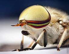 The male Tabanus lineola has holoptic compound eyes, with the dorsal ommatidia larger than the ventral ommatidia
 -
 In some male mayflies the eyes are split into separate organs for distinct visual functions
 -
 Stalked dichoptic eyes of a River Crab are typical of mature larger Crustacea. The reflection of the photographer in different regions of the surface of each eye indicate the basis for stereoscopic vision
 -
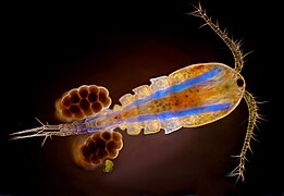 Many copepods and other small Crustacea have their eyes combined into a single cycloptic compound eye in the middle of the head, visible here as the dark spot between the bases of the antennae
.png)
.jpg)


Most species of Arthropoda with compound eyes bear just two eyes that are located separately and symmetrically, one on each side of the head. This arrangement is called dichoptic. Examples include most insects, and most of the larger species of Crustacea, such as crabs. Many other organisms, such as vertebrates and Cephalopoda are similarly and analogously dichoptic, which is the common state in animals that are members of the
On the other hand, some modes of life demand enhanced
In contrast, the need for particular functions may not require extremely large eyes, but do require great resolution and good stereoscopic vision for precise attacks. Good examples may be seen in the Mantodea and Mantispidae, in which seeing prey from particular ommatidia in both compound eyes at the same time, indicates that it is in the right position to snatch in a close-range ambush. Their eyes accordingly are placed in a good position for all-round vision, plus particular concentration on the anterior median plane. The individual ommatidia are directed in all directions and accordingly, one may see a dark spot (the pseudopupil), showing which ommatidia are covering that field of view; from any position on the median plane, and nowhere else, the two dark spots are symmetrical and identical.
Sometimes the needs for visual acuity in different functions conflict, and different parts of the eyes may be adapted to separate functions; for example, the Gyrinidae spend most of their adult lives on the surface of water, and have their two compound eyes split into four halves, two for underwater vision and two for vision in air. Again, particularly in some Diptera, ommatidia in different regions of the holoptic male eye may differ visibly in size; the upper ommatidia tend to be larger. In the case of some Ephemeroptera the effect is so exaggerated that the upper part of the eye is elevated like a risen cupcake, while its lower part that serves for routine vision looks like a separate organ.
Compound eyes are often not completely symmetrical in terms of ommatidia count. For example, asymmetries have been indicated in honeybees[6] and various flies.[7] This asymmetry has been correlated with behavioural lateralization in ants (turning bias).[8]
Genetic controls
In the fruit fly
The visual systems of Chelicerata (the sister group to the remaining Arthropoda) are less well understood. It has been shown that homologs of many eye patterning genes are variably expressed in the eyes of different spider species, but the functional significance of these changes in expression is not well understood, due to lack of functional data.[13][14] In addition, it has been shown in horseshoe crabs and spiders that Pax6 homologs are not expressed in the same way as their counterparts in insects, suggesting that Pax6 may not be required as a top-level eye patterning switch in chelicerates.[15][16] Most of the functional data on eye patterning in Chelicerata is drawn from the daddy-longlegs Phalangium opilio, which has been used to show that eyes absent plays a conserved role in patterning both the visual systems of this species (an example of conservation of gene function, with respect to insects) and that dachshund affects the patterning of lateral eyes, but not median eyes (another example of conservation).[17]
Evolution
Hexapods are currently thought to fall within the Crustacean crown group; while molecular work paved the way for this association, their eye morphology and development is also markedly similar.[18] The eyes are strikingly different from the myriapods, which were traditionally considered to be a sister group to the Hexapoda.
Both ocelli and compound eyes were probably present in the last common arthropod ancestor,
Origin
No fossil organisms have been identified as similar to the last common ancestor of arthropods; hence the eyes possessed by the first arthropod remains a matter of conjecture. The largest clue into their appearance comes from the onychophorans: a stem group lineage that diverged soon before the first true arthropods. The eyes of these creatures are attached to the brain using nerves which enter into the centre of the brain, and there is only one area of the brain devoted to vision. This is similar to the wiring of the median ocelli (small simple eyes) possessed by many arthropods; the eyes also follow a similar pathway through the early development of organisms. This suggests that onychophoran eyes are derived from simple ocelli, and the absence of other eye structures implies that the ancestral arthropod lacked compound eyes, and only used median ocelli to sense light and dark.[2]

A conflicting view notes, however, that compound eyes appeared in many early arthropods, including the trilobites and eurypterids. That suggests that the compound eye may have developed after the onychophoran and arthropod lineages split, but before the radiation of arthropods.[20] This view is supported if a stem-arthropod position is supported for compound-eye bearing Cambrian organisms such as the Radiodontids. Yet another alternative is that compound eyes independently evolved, multiple times within the arthropods.[21]
There were probably only a single pair of ocelli in the arthropod concestor, since Cambrian lobopod fossils display a single pair. And while many arthropods today have three, four, or even six, the lack of a common pathway suggests that a pair is the most probable ancestral state. The crustaceans and insects mainly have three ocelli, suggesting that such a formation was present in their concestor.[2]
It is deemed probable that the compound eye arose as a result of the 'duplication' of individual ocelli.[20] In turn, the dispersal of compound eyes seems to have created large networks of seemingly independent eyes in some arthropods, such as the larvae of certain insects.[20] In some other insects and myriapods, lateral ocelli appear to have arisen by the reduction of lateral compound eyes.[20]
Trilobite eyes
The eyes of trilobites came in three forms, called holochroal, schizochroal, and abathochroal eyes. The eye morphology of trilobites is useful for inferring their mode of life, and can function as indicators of the palaeo-environment conditions.[22]
The
The more complex schizochroal eye was found only in one sub-order of trilobite, the Phacopina (Ordovician-Silurian). There is no exact counterpart to the schizochroal eye in modern animals, but a somewhat similar eye structure is found in adult male insects in the order Strepsiptera. Schizochroal eyes developed as an improvement on holochroal; they were more powerful, with overlapping visual fields, and were particularly useful for nocturnal vision and possibly for colour and depth perception. Schizochroal eyes have up to 700 large lenses (large compared to holochroal lenses). Each lens has a cornea, and each has an individual sclera that separates it from the surrounding lenses. The multiple lenses for the eye were each constructed from a single calcite crystal. Early schizochroal eye designs appear haphazard and irregular – possibly constrained by the geometrical complications of packing identical sized lenses on a curved surface. Later schizochroal eyes had size graduated lens.[22]
The abathochroal eye is the third eye morphology of trilobites, but it has found only within the Eodiscina. This form of eye consisted of up to 70 much smaller lenses. The cornea separated each lens, and the sclera on each lens terminated on top of each cornea.[22]
Horseshoe crab

The
Horseshoe crabs have two large compound eyes on the sides of its head. An additional simple eye is positioned at the rear of each of these structures.[24] In addition to these obvious structures, it also has two smaller ocelli situated in the middle-front of its carapace, which may superficially be mistaken for nostrils.[24] A further simple eye is located beneath these, on the underside of the carapace; this eye is initially paired during embryonic stages and fuses later in development.[24][15] A further pair of simple eyes are positioned just in front of the mouth.[24] The simple eyes are probably important during the embryonic or larval stages of the organism, with the compound eyes and median ocelli becoming the dominant sight organs during adulthood.[24] These ocelli are less complex, and probably less derived, than those of the Mandibulata.[20] Unlike the compound eyes of trilobites, those of horseshoe crabs are triangular in shape; they also have a generative region at their base, but this elongates with time. Hence the one ommatidium at the apex of the triangle was the original "eye" of the larval organism, with subsequent rows added as the organism grew.[18]
Insects and crustaceans

It is generally thought that
Although stalked eyes on peduncles occur in some species of crustaceans and some insects, only some of the Crustacea, such as crabs, bear their eyes on articulated peduncles that permit the eyes to be folded out of the way of trouble.
Myriapods

Most myriapods bear stemmata – single lensed eyes which are thought to have evolved by the reduction of a compound eye.[20] However, members of the chilopod genus Scutigera have a compound eye, which is composed of facets [26] and not, as earlier interpretations had it, of clustered stemmata.[21] that were thought to grow in rows, inserted between existing rows of ocelli.[18]
See also
Footnotes
- Ocelliare about 5000 times more sensitive than apposition compound eyes. They can, for instance, respond to the position of the full moon.
References
- ^ a b
Land, Michael F.; Fernald, Russell D. (1992), "The Evolution of Eyes", Annual Review of Neuroscience, 15 (1): 1–29, PMID 1575438.
- ^ a b c
Mayer, G. (2006). "Structure and development of onychophoran eyes: What is the ancestral visual organ in arthropods?". Arthropod Structure & Development. 35 (4): 231–245. PMID 18089073.
- ^ a b c Taylor, Charles P. (1981). "Contribution of compound eyes and ocelli to steering of locusts in flight. I. Behavioural analysis". J. Exp. Biol. 93: 1–18. ]
- ^
Meyer-Rochow, V.B. (1974). "Structure and function of the larval eye of the sawfly larva Perga". Journal of Insect Physiology. 20 (8): 1565–1591. PMID 4854430.
- ^
Wilson, M. (1978). "The functional organisation of locust ocelli". Journal of Comparative Physiology. 124 (4): 297–316. S2CID 572458.
- ^
Seidl, R.; Kaiser, W. (1981). "Visual field size, binocular domain and the ommatidial array of the compound eyes in worker honey bees". J. Comp. Physiol. 143: 17–26. S2CID 41150125.
- ^
Beersma, D.G.; et al. (1977). "Retinal lattice, visual field and binocularities in flies". J. Comp. Physiol. 119 (3): 207–220. S2CID 6384399.
- ^
Hunt, E.R.; et al. (2018). "Asymmetric ommatidia count and behavioural lateralization in the ant Temnothorax albipennis". PMID 29643429.
- PMID 17618111.
- PMID 8582270
- PMID 7821215.
- S2CID 4248264
- PMID 26034574.
- PMID 26034575.
- ^ PMID 18651657.
- S2CID 253246397.
- S2CID 267821504.
- ^ PMID 18089079
- PMID 18089081
- ^ ISBN 978-0-8493-3498-6
- ^
- ^ ISBN 9781444313321– via Google Books.
- ^
Barlow, R.B. (2009). "Vision in horseshoe crabs". In Tanacredi, J.T.; Botton, M.L.; Smith, D. (eds.). Biology and Conservation of Horseshoe Crabs. Springer. pp. 223–235. ISBN 9780387899589– via Google Books.
- ^ PMID 18089075.
- S2CID 89428386.
- S2CID 6466405.
Vision in animals | ||
|---|---|---|
| Vision | .jpg) | |
| Eyes |
| |
| Evolution | ||
| Coloration | ||
| Related topics | ||