
Astrogliosis
| Astrogliosis | |
|---|---|
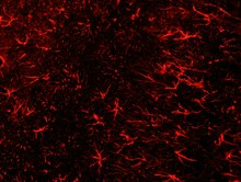
 Formation of reactive astrocytes after central nervous system (CNS) injury | |
| Anatomical terminology |
Astrogliosis (also known as astrocytosis or referred to as reactive astrogliosis) is an abnormal increase in the number of
Causes
Reactive astrogliosis is a spectrum of changes in
Insults to neurons in the central nervous system caused by infection, trauma, ischemia, stroke, recurring seizures, autoimmune responses, or other neurodegenerative diseases may cause reactive astrocytes.[2]
When the astrogliosis is pathological itself, instead of a normal response to a pathological problem, it is referred to as astrocytopathy.[5]
Functions and effects
Reactive astrocytes may benefit or harm surrounding neural and non-neural cells. They undergo a series of changes that may alter astrocyte activities through gain or loss of functions lending to neural protection and repair,
Neural protection and repair
Proliferating reactive astrocytes are critical to scar formation and function to reduce the spread and persistence of inflammatory cells, to maintain the repair of the blood–brain barrier (BBB), to decrease tissue damage and lesion size, and to decrease neuronal loss and demyelination.[6][7]
Reactive astrocytes defend against oxidative stress through glutathione production and have the responsibility of protecting CNS cells from NH4+ toxicity.[3]
They protect CNS cells and tissue through various methods,
They have also been shown to reduce
Scar formation
Proliferating reactive scar-forming astrocytes are consistently found along borders between healthy tissues and pockets of damaged tissue and inflammatory cells. This is usually found after a rapid, locally triggered inflammatory response to acute traumatic injury in the spinal cord and brain. In its extreme form, reactive astrogliosis can lead to the appearance of newly proliferated astrocytes and scar formation in response to severe tissue damage or inflammation.
Molecular triggers that lead to this scar formation include
Regulation of inflammation
Reactive astrocytes are related to the normal function of astrocytes. Astrocytes are involved in the complex regulation of CNS inflammation that is likely to be context-dependent and regulated by multimodal extra- and intracellular signaling events. They have the capacity to make different types of molecules with either pro- or anti-inflammatory potential in response to different types of stimulation. Astrocytes interact extensively with microglia and play a key role in CNS inflammation. Reactive astrocytes can then lead to abnormal function of astrocytes and affect their regulation and response to inflammation.[10][11]
Pertaining to anti-inflammatory effects, reactive scar-forming astrocytes help reduce the spread of inflammatory cells during locally initiated inflammatory responses to traumatic injury or during peripherally-initiated adaptive immune responses.[3][8] In regard to pro-inflammatory potential, certain molecules in astrocytes are associated with an increase in inflammation after traumatic injury.[3]
At early stages after insults, astrocytes not only activate inflammation, but also form potent cell migration barriers over time. These barriers mark areas where intense inflammation is needed and restrict the spread of inflammatory cells and infectious agents to nearby healthy tissue.[3][7][8] CNS injury responses have favored mechanisms that keep small injuries uninfected. Inhibition of the migration of inflammatory cells and infectious agents have led to the accidental byproduct of axon regeneration inhibition, owing to the redundancy between migration cues across cell types.[3][7]
Biological mechanisms
Changes resulting from astrogliosis are regulated in a context-specific manner by specific signaling events that have the potential to modify both the nature and degree of these changes. Under different conditions of stimulation,
Signaling molecules
Few of the known signaling molecules and their effects are understood in the context of reactive astrocytes responding to different degrees of insult.
Upregulation of
Transporters and channels
The presence of astrocyte
Neurological pathologies
Loss or disturbance of functions normally performed by
- Autoimmune destruction of astrocyte endfeet that contact and envelop blood vessels is associated with CNS inflammation and a form of multiple sclerosis
- seizures
- In seizures, psychomotordisturbances, and premature death.
- In a familial form of amyotropic lateral sclerosis (ALS), a dominant gain-of-function mutation of the gene encoding superoxide dismutase(SOD) leads to production of reactive astrocytes of molecules that are toxic to motor neurons.
Reactive astrocytes may also be stimulated by specific signaling cascades to gain detrimental effects such as the following:[3][14]
- Exacerbation of inflammation via cytokine production
- Production and release of neurotoxic levels of reactive oxygen species
- Release of potentially excitotoxic glutamate
- The potential contribution to seizure genesis
- Compromise of endothelial growth factorproduction
- Cytotoxic AQP4overactivity
- Potential for chronic astrocytesto contribute to chronic pain
Reactive astrocytes have the potential to promote neural toxicity via the generation cytotoxic molecules such as nitric oxide radicals and other reactive oxygen species,[7] which may damage nearby neurons. Reactive astrocytes may also promote secondary degeneration after CNS injury.[7]
Novel therapeutic techniques
Due to the destructive effects of astrogliosis, which include altered molecular expression, release of inflammatory factors, astrocyte proliferation and neuronal dysfunction, researchers are currently searching for new ways to treat astrogliosis and neurodegenerative diseases. Various studies have shown the role of astrocytes in diseases such as
Anti-gliosis function of BB14
One specific drug candidate is BB14, which is a nerve growth factor-like peptide that acts as a
Regulatory function of TGFB
Ethidium bromide treatment
Injection of
Metalloprotinease activity
Oligodendrocyte precursor cells and C6