
Axillary artery
| Axillary artery | |
|---|---|
Posterior circumflex humeral continues as brachial artery | |
| Vein | axillary vein |
| Supplies | axilla |
| Identifiers | |
| Latin | arteria axillaris |
| MeSH | D001366 |
| TA98 | A12.2.09.002 |
| TA2 | 4616 |
| FMA | 22654 |
| Anatomical terminology] | |
In
After passing the lower margin of teres major it becomes the brachial artery.
Structure
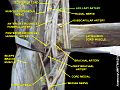
The axillary artery is often referred to as having three parts, with these divisions based on its location relative to the Pectoralis minor muscle, which is superficial to the artery.
- First part – the part of the artery superior to the pectoralis minor
- Second part – the part of the artery posterior to the pectoralis minor
- Third part – the part of the artery inferior to the pectoralis minor.
Relations
The axillary artery is accompanied by the axillary vein,[1] which lies medial to the artery, along its length.
In the axilla, the axillary artery is surrounded by the brachial plexus.[1] The second part of the axillary artery is the reference for the locational descriptions of the cords in the brachial plexus. For example, the posterior cord of the brachial plexus is so named because it lies posterior to the second part of the artery.
Branches

The axillary artery has several smaller branches. The branches can be remembered, in order, when traveling from the heart, with the
- First part (1 branch)
- Superior thoracic artery (Supreme thoracic artery)[1]
- Second part (2 branches)
- Thoraco-acromial artery[1]
- Lateral thoracic artery.[1] If the lateral thoracic artery is not branching from the axillary artery, will most likely branch from the following (in order of likelihood): (1) thoracoacromial, (2) third part of axillary artery, (3) suprascapular artery, (4) subscapular artery
- Third part (3 branches)
- Subscapular artery[1]
- Anterior humeral circumflex artery[1]
- Posterior humeral circumflex artery[1]
Continues as the
Clinical significance
The axillary artery can be safely clamped without endangering the arm, but only in a location proximal to the origin of the subscapular artery (and distal to the thyrocervical trunk of the subclavian artery). The anastomotic network surrounding the scapula provides an alternate path for collateral circulation to the arm from arteries including the
The right axillary artery is often used as an arterial cannulation site in cardiac surgery, particularly for repair of aortic dissection and replacement of the ascending aorta and aortic arch.
Additional images
-
 The veins of the right axilla, viewed from in front.
 -
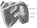
 The right brachial plexus (infraclavicular portion) in the axillary fossa; viewed from below and in front.
 -
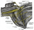
 Suprascapular and axillary nerves of right side, seen from behind.
 -
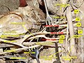 Brachial plexus and axillary artery
 -
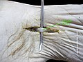 Axillary artery
 -
 Axillary artery
 -
 Axillary artery
 -
 Axillary artery
 -
 Axillary artery
 -
 Axillary artery


References
- ^ ISBN 9780702029714.
- ^ MedicalMnemonics.com: 1208 852 663
External links
- lesson3axillaryart&vein at The Anatomy Lesson by Wesley Norman (Georgetown University)
- Axillary artery at the Duke University Health System's Orthopedics program
- Anatomy photo:05:06-0101 at the SUNY Downstate Medical Center – "Axillary Region: Parts of the Axillary Artery"
- Anatomy figure: 05:04-01 at Human Anatomy Online, SUNY Downstate Medical Center – "The axillary artery and its major branches shown in relation to major landmarks."