Batrachochytrium dendrobatidis
| Batrachochytrium dendrobatidis | |
|---|---|

| |
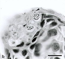
Zoosporangia of B. dendrobatidis growing on a freshwater arthropod (a) and algae (b); scale bars = 30 μm
| |
| Scientific classification | |
| Domain: | Eukaryota |
| Kingdom: | Fungi |
| Division: | Chytridiomycota |
| Class: | Chytridiomycetes |
| Order: | Rhizophydiales |
| Family: | Batrachochytriaceae |
| Genus: | Batrachochytrium |
| Species: | B. dendrobatidis
|
| Binomial name | |
| Batrachochytrium dendrobatidis Longcore, Pessier & D.K. Nichols (1999)
| |
Batrachochytrium dendrobatidis (
Since its discovery in 1998 by
The fungal pathogens that cause the disease chytridiomycosis ravage the skin of frogs, toads, and other amphibians, throwing off their balance of water and salt and eventually causing heart failure,
Etymology
The generic name is derived from the Greek words batrachos (frog) and chytra (earthen pot), while the
Systematics
Batrachochytrium dendrobatidis was until recently considered the single species of the genus Batrachochytrium. The initial classification of the pathogen as a chytrid was based on zoospore ultrastructure.
Morphology
B. dendrobatidis infects the
Zoospore structure
Flagellum structure
A nonfunctioning
Life cycle

B. dendrobatidis has two primary life stages: a sessile, reproductive
Besides amphibians B. dendrobatidis also infects crayfish (
Physiology
B. dendrobatidis can grow within a wide temperature range (4-25 °C), with optimal temperatures being between 17 and 25 °C.
Varying forms
B. dendrobatidis has occasionally been found in forms distinct from its traditional zoospore and sporangia stages. For example, before the
Habitat and relationship to amphibians
The fungus grows on amphibian skin and produces aquatic zoospores.
Geographic distribution
It has been suggested that B. dendrobatidis originated in Africa or Asia and subsequently spread to other parts of the world by trade in African clawed frogs (Xenopus laevis).[16] In this study, 697 archived specimens of three species of Xenopus, previously collected from 1879 to 1999 in southern Africa, were examined. The earliest case of chytridiomycosis was found in a X. laevis specimen from 1938. The study also suggests that chytridiomycosis had been a stable infection in southern Africa from 23 years prior to finding any infected outside of Africa.[16] There is more recent information that the species originated on the Korean peninsula and was spread by the trade in frogs.[17]
American bullfrogs (
A wide variety of amphibian hosts have been identified as being susceptible to infection by B. dendrobatidis, including wood frogs (
Southeast Asia
While most studies concerning B. dendrobatidis have been performed in various locations across the world, the presence of the fungus in Southeast Asia remains a relatively recent development. The exact process through which the fungus was introduced to Asia is not known, however, as mentioned above, it has been suggested transportation of
In Cambodia, a study showed B. dendrobatidis to be prevalent throughout the country in areas near
Effect on amphibians
Worldwide amphibian populations have been on a steady decline due to an increase in the disease Chytridiomycosis, caused by this Bd fungus. Bd can be introduced to an amphibian primarily through water exposure, colonizing the digits and ventral surfaces of the animal's body most heavily and spreading throughout the body as the animal matures. Potential effects of this pathogen are hyperkeratosis, epidermal hyperplasia, ulcers, and most prominently the change in osmotic regulation often leading to cardiac arrest.[44] The death toll on amphibians is dependent on a variety of factors but most crucially on the intensity of infection. Certain frogs adopt skin sloughing as a defense mechanism for B. dendrobatidis; however, this is not always effective, as mortality fluctuates between species. For example, the Fletcher frog, despite practising skin sloughing, suffers from a particularly high mortality rate when infected with the disease compared to similar species like Lim. peronii and Lim. tasmaniensis. Some amphibian species have been found to adapt to infection after an initial die-off with survival rates of infected and non-infected individuals being equal.[45]
According to a study by the Australian National University estimates that the Bd fungus has caused the decline of 501 amphibian species—about 6.5 percent of the world known total. Of these, 90 have been entirely wiped out and another 124 species have declined by more than 90 percent, and their odds of the effected species recovering to a healthy population are doubtful.[46] However, these conclusions were criticized by later studies, which proposed that Bd was not as primary a driver of amphibian declines as found by the previous study.[47]
One amphibian in particular that Batrachochytrium dendrobatidis (Bd) has affected greatly was the Lithobates clamitans. Bd kills this frog by interfering with external water exchange thereby causing an imbalance with ion exchange which leads to heart failure.
Immunity
Some amphibian species are actually immune to Bd, or have biological protections against the fungus.[48] One such salamander is the alpine salamander, or S. atra. These salamanders have several subspecies, but they share a common trait: toxicity. A 2012 study demonstrated that no alpine salamanders in the area had the disease, despite its prevalence in the area.[49] Alpine salamanders can produce alkaloid products[50][49] or other toxic peptides[50] that may be protective against microbes.[51]
See also
- Pathogenic fungi
- Decline in amphibian populations
- Ranavirus
References
- PMID 9671799.
- ^ JSTOR 3761366.
- PMC 4918143.
- ^ PMID 24003137.
- ^ PMID 17148429.[permanent dead link]
- PMID 18488347.
- S2CID 8545084.
- PMID 16465834.
- S2CID 52850132.
- PMID 23248288.
- ^ PMID 21148822.
- PMID 12887256.
- S2CID 4421285.
- S2CID 84272576.
- S2CID 16838374.
- ^ PMID 15663845.
- ISSN 0362-4331. Retrieved 2018-05-20.
- S2CID 85969277.
- ^ Daszak P, Strieby A, Cunningham AA, Longcore JE, Brown CC, Porter D (2004). "Experimental evidence that the bullfrog (Rana catesbeiana) is a potential carrier of chytridiomycosis, an emerging fungal disease of amphibians". Herpetological Journal. 14: 201–207.
- PMID 16119886.
- S2CID 86205459.
- S2CID 16838374. Archived from the original(PDF) on 2008-12-26.
- PMID 15997823.
- S2CID 11114174.
- ^ Reeves MK (2008). "Batrachochytrium dendrobatidis in wood frogs (Lithobates sylvatica) from Three National Wildlife Refuges in Alaska, USA". Herpetological Review. 39 (1): 68–70.
- PMID 18689660.
- S2CID 84713825.
- S2CID 23997421.
- ^ Brodman R, Briggler JT (2008). "Batrachochytrium dendrobatidis in Ambystoma jeffersonianum larvae in southern Indiana". Herpetological Review. 39 (3): 320–321.
- ^ a b Lehtinen RM, Kam Y-C Richards CL (2008). "Preliminary surveys for Batrachochytrium dendrobatidis in Taiwan". Herpetological Review. 39 (3): 317–318.
- ^ Lovich R, Ryan MJ, Pessier AP, CLaypool B (2008). "Infection with the fungus Batrachochytrium dendrobatidis in a non-native Lithobates berlandieri below sea level in the Coachella Valley, California, USA". Herpetological Review. 39 (3): 315–317.
- PMID 18689659.
- PMID 25266902.
- S2CID 24321977.
- PMID 18286805.
- PMID 19244970.
- PMID 19575560.
- ^ McLeod DS, Sheridan JA, Jiraungkoorskul W, Khonsue W (2008). "A survey for chytrid fungus in Thai amphibians". Raffles Bulletin of Zoology. 56: 199–204.
- PMID 19899344.
- PMID 21268987.
- ^ Gaertner JP, Mendoza JA, Forstner MR, Neang T, Hahn D (2011). "Detection of Batrachochytrium dendrobatidis in frogs from different locations in Cambodia". Herpetological Review. 42: 546–548.
- ^ Mendoza JA, Gaertner JP, Holden J, Forstner MR, Hahn D (2011). "Detection of Batrachochytrium dendrobatidis on amphibians in Pursat Province, Cambodia". Herpetological Review. 42: 542–545.
- PMID 25902035.
- ^ "Chytridiomycosis". www.amphibiaweb.org. Retrieved 2016-05-27.
- PMID 30368999.
- ^ Yong, Ed (2019-03-28). "The Worst Disease Ever Recorded". The Atlantic. Retrieved 2019-03-28.
- PMID 32193293.
- S2CID 232030040.
- ^ OCLC 1030045649.)
{{cite book}}: CS1 maint: multiple names: authors list (link - ^ S2CID 221862886.
- S2CID 253637272.
Further reading
- Daszak, Peter; Berger L; Cunningham AA; Hyatt AD; Green DE; Speare R. (1999). "Emerging Infectious Diseases and Amphibian Population Declines". Emerging Infectious Diseases. 5 (6): 735–748. PMID 10603206.
- Johnson, Megan L.; Speare, Richard (August 2003). "Survival of Batrachochytrium dendrobatidis in Water: Quarantine and Disease Control Implications". Emerging Infectious Diseases. 9 (8): 915–921. PMID 12967488.
