
Beta sheet

The beta sheet (β-sheet, also β-pleated sheet) is a common
History

The first β-sheet structure was proposed by
Structure and orientation
Geometry
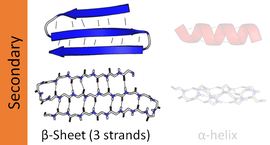
The majority of β-strands are arranged adjacent to other strands and form an extensive

However, β-strands are rarely perfectly extended; rather, they exhibit a twist. The energetically preferred
The side chains point outwards from the folds of the pleats, roughly perpendicularly to the plane of the sheet; successive amino acid residues point outwards on alternating faces of the sheet.
Hydrogen bonding patterns
Because peptide chains have a directionality conferred by their
In an antiparallel arrangement, the successive β-strands alternate directions so that the N-terminus of one strand is adjacent to the C-terminus of the next. This is the arrangement that produces the strongest inter-strand stability because it allows the inter-strand hydrogen bonds between carbonyls and amines to be planar, which is their preferred orientation. The peptide backbone dihedral angles (φ, ψ) are about (–140°, 135°) in antiparallel sheets. In this case, if two atoms Cα
i and Cα
j are adjacent in two hydrogen-bonded β-strands, then they form two mutual backbone hydrogen bonds to each other's flanking peptide groups; this is known as a close pair of hydrogen bonds.
In a parallel arrangement, all of the N-termini of successive strands are oriented in the same direction; this orientation may be slightly less stable because it introduces nonplanarity in the inter-strand hydrogen bonding pattern. The dihedral angles (φ, ψ) are about (–120°, 115°) in parallel sheets. It is rare to find less than five interacting parallel strands in a motif, suggesting that a smaller number of strands may be unstable, however it is also fundamentally more difficult for parallel β-sheets to form because strands with N and C termini aligned necessarily must be very distant in sequence [citation needed]. There is also evidence that parallel β-sheet may be more stable since small amyloidogenic sequences appear to generally aggregate into β-sheet fibrils composed of primarily parallel β-sheet strands, where one would expect anti-parallel fibrils if anti-parallel were more stable.
In parallel β-sheet structure, if two atoms Cα
i and Cα
j are adjacent in two hydrogen-bonded β-strands, then they do not hydrogen bond to each other; rather, one residue forms hydrogen bonds to the residues that flank the other (but not vice versa). For example, residue i may form hydrogen bonds to residues j − 1 and j + 1; this is known as a wide pair of hydrogen bonds. By contrast, residue j may hydrogen-bond to different residues altogether, or to none at all.
The hydrogen bond arrangement in parallel beta sheet resembles that in an amide ring motif with 11 atoms.
Finally, an individual strand may exhibit a mixed bonding pattern, with a parallel strand on one side and an antiparallel strand on the other. Such arrangements are less common than a random distribution of orientations would suggest, suggesting that this pattern is less stable than the anti-parallel arrangement, however bioinformatic analysis always struggles with extracting structural thermodynamics since there are always numerous other structural features present in whole proteins. Also proteins are inherently constrained by folding kinetics as well as folding thermodynamics, so one must always be careful in concluding stability from bioinformatic analysis.
The hydrogen bonding of β-strands need not be perfect, but can exhibit localized disruptions known as β-bulges.
The hydrogen bonds lie roughly in the plane of the sheet, with the
Amino acid propensities
Large aromatic residues (tyrosine, phenylalanine, tryptophan) and β-branched amino acids (threonine, valine, isoleucine) are favored to be found in β-strands in the middle of β-sheets. Different types of residues (such as proline) are likely to be found in the edge strands in β-sheets, presumably to avoid the "edge-to-edge" association between proteins that might lead to aggregation and amyloid formation.[2]
Common structural motifs


β-hairpin motif
A very simple structural motif involving β-sheets is the β-hairpin, in which two antiparallel strands are linked by a short loop of two to five residues, of which one is frequently a glycine or a proline, both of which can assume the dihedral-angle conformations required for a tight turn or a β-bulge loop. Individual strands can also be linked in more elaborate ways with longer loops that may contain α-helices.
Greek key motif
The Greek key motif consists of four adjacent antiparallel strands and their linking loops. It consists of three antiparallel strands connected by hairpins, while the fourth is adjacent to the first and linked to the third by a longer loop. This type of structure forms easily during the protein folding process.[3][4] It was named after a pattern common to Greek ornamental artwork (see meander).
β-α-β motif
Due to the chirality of their component amino acids, all strands exhibit right-handed twist evident in most higher-order β-sheet structures. In particular, the linking loop between two parallel strands almost always has a right-handed crossover chirality, which is strongly favored by the inherent twist of the sheet.


β-meander motif
A simple
The vast majority of β-meander regions in proteins are found packed against other motifs or sections of the polypeptide chain, forming portions of the hydrophobic core that canonically drives formation of the folded structure.[9] However, several notable exceptions include the Outer Surface Protein A (OspA) variants[6] and the Single Layer β-sheet Proteins (SLBPs)[10] which contain single-layer β-sheets in the absence of a traditional hydrophobic core. These β-rich proteins feature an extended single-layer β-meander β-sheets that are primarily stabilized via inter-β-strand interactions and hydrophobic interactions present in the turn regions connecting individual strands.
Psi-loop motif
The psi-loop (Ψ-loop) motif consists of two antiparallel strands with one strand in between that is connected to both by hydrogen bonds.
Structural architectures of proteins with β-sheets
β-sheets are present in
Structural topology
The topology of a β-sheet describes the order of
β-sheets can be open, meaning that they have two edge strands (as in the
Dynamic features
β-pleated sheet structures are made from extended β-strand polypeptide chains, with strands linked to their neighbours by
Parallel β-helices

A β-helix is formed from repeating structural units consisting of two or three short β-strands linked by short loops. These units "stack" atop one another in a helical fashion so that successive repetitions of the same strand hydrogen-bond with each other in a parallel orientation. See the β-helix article for further information.
In lefthanded β-helices, the strands themselves are quite straight and untwisted; the resulting helical surfaces are nearly flat, forming a regular triangular prism shape, as shown for the 1QRE archaeal carbonic anhydrase at right. Other examples are the lipid A synthesis enzyme LpxA and insect antifreeze proteins with a regular array of Thr sidechains on one face that mimic the structure of ice.[17]

Righthanded β-helices, typified by the pectate lyase enzyme shown at left or P22 phage tailspike protein, have a less regular cross-section, longer and indented on one of the sides; of the three linker loops, one is consistently just two residues long and the others are variable, often elaborated to form a binding or active site.[18]
A two-sided β-helix (right-handed) is found in some bacterial
In pathology
Some proteins that are disordered or helical as monomers, such as amyloid β (see
The side chains from the amino acid residues found in a β-sheet structure may also be arranged such that many of the adjacent sidechains on one side of the sheet are hydrophobic, while many of those adjacent to each other on the alternate side of the sheet are polar or charged (hydrophilic),[21] which can be useful if the sheet is to form a boundary between polar/watery and nonpolar/greasy environments.
See also
- Collagen helix
- Foldamers
- Folding (chemistry)
- Tertiary structure
- α-helix
- Structural motif
References
- ISBN 0-471-19350-X.
- PMID 11880627.
- ^ Tertiary Protein Structure and Folds: section 4.3.2.1. From Principles of Protein Structure, Comparative Protein Modelling, and Visualisation
- PMID 8506258.
- )
- ^ PMID 17093048.
- ^ "SCOP: Fold: WW domain-like". Archived from the original on 2012-02-04. Retrieved 2007-06-01.
- ^ PPS '96 – Super Secondary Structure
- PMID 20133689.
- PMID 31306512.
- PMID 8745398.
- ^ S2CID 28921557.
- PMID 9016544.
- PMID 24304899.
- PMID 7115900.
- PMID 4052563.
- S2CID 4385352.
- ISBN 0-8153-2305-0.
- PMID 8253063.
- PMID 15944695.
- PMID 7682699.
Further reading
- Cooper J (31 May 1996). "Super Secondary Structure - Part II". Principles of Protein Structure Using the Internet. Retrieved 25 May 2007.
- "Open-sided Beta-meander". Structural Classification of Proteins (SCOP). 20 October 2006. Archived from the original on 4 February 2012. Retrieved 31 May 2007.