Bicornuate uterus
| Bicornuate uterus | |
|---|---|
| Other names | bicornate uterus |
 | |
| Two types of a human bicornuate uterus | |
| Specialty | Gynaecology |
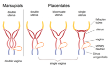
A bicornuate uterus or bicornate uterus (from the Latin cornū, meaning "horn"), is a type of müllerian anomaly in the human uterus, where there is a deep indentation at the fundus (top) of the uterus.
Pathophysiology
A bicornuate uterus develops during
Diagnosis
Diagnosis of bicornuate uterus typically involves imaging of the uterus with 2D or 3D ultrasound, hysterosalpingography, or magnetic resonance imaging (MRI). On imaging, a bicornuate uterus can be distinguished from a septate uterus by the angle between the cornua (intercornual angle): less than 75 degrees in a septate uterus, and greater than 105 degrees in a bicornuate uterus. Measuring the depth of the cleft between the cornua (fundal cleft) may also assist in diagnosis; a cleft of over 1 centimetre (0.39 in) is indicative of bicornuate uterus.[2]
Classification
Bicornuate uterus is typically classified based on whether or not the division extends to the external cervical os. Bicornuate uteri with a division above the os are called bicornuate unicollis and those with a divided os are called bicornuate bicollis.
There is also a hybrid bicornuate uterus: External fundal depressions of variable depths associated with a septate uterus can be seen by laparoscopy, indicating the coexistence of the two anomalies. These cases are candidates for hysteroscopic metroplasty under appropriate sonographic and/or laparoscopic monitoring.[4]
An obstructed bicornuate uterus showing uni or bilateral obstruction might also be possible. The unilateral obstruction is more difficult to diagnose than the bilateral obstructive. A delay in the diagnosis can be problematic and compromise the reproductive abilities of those cases.[5]
Treatment
Bicornuate uterus typically requires no treatment.[1] In those who do need treatment, metroplasty is the surgical correction of choice.[2] Women who have recurrent miscarriage with no other explanation may benefit from surgery.[6]
Epidemiology
The occurrence of all types of paramesonephric duct abnormalities in women is estimated around 0.4%.[7] A bicornuate uterus is estimated to occur in 0.1–0.6% of women in the US.[8] It is possible that this figure is an underestimate, since subtle abnormalities often go undetected.[citation needed] Some intersex individuals whose external genitalia are perceived as being male may nonetheless have a variably shaped uterus. Women exposed in utero to diethylstilbestrol (DES) are at risk for this abnormality.[citation needed]
In pregnancy
A bicornuate uterus is an indication for increased surveillance of a pregnancy, though most women with a bicornuate uterus are able to have healthy pregnancies.
Fetuses developing in bicornuate uteri are more likely to present breech or transverse, with the fetal head in one horn and the feet in the other. This will often necessitate cesarean delivery. If the fetus is vertex (head down), the two horns may not contract in coordination, or the horn that does not contain the pregnancy may interfere with contractions and descent of the fetus, causing obstructed labor.[13]
Effect on intrauterine device usage
Usage of
Uterus bicornis in mammals

A double uterus is the normal female anatomy in many
Health issues
The risk of cancer is not increased in the case of uterus bicornis.
References
- ^ a b c Bauman, D. (2013). "Pediatric & Adolescent Gynecology". CURRENT Diagnosis & Treatment: Obstetrics & Gynecology. McGraw-Hill.
- ^ a b c d e f Cunningham F, Leveno KJ, Bloom SL, Dashe JS, Hoffman BL, Casey BM, Spong CY (eds.). "Congenital Genitourinary Abnormalities". Williams Obstetrics (25 ed.). McGraw-Hill.
- PMID 18367185.
- S2CID 6177612.
- PMID 21514191.
- ^ Hoffman BL, Schorge JO, Bradshaw KD, Halvorson LM, Schaffer JI, Corton MM (eds.). "Anatomic Disorders". Williams Gynecology (3 ed.).
- PMID 10982475.
- ^ "Bicornuate Uterus - an overview | ScienceDirect Topics". www.sciencedirect.com. Retrieved 2021-01-04.
- S2CID 5476966.
- S2CID 22903707.
- S2CID 72723061.
- PMID 9521976.
- ^ a b c Pascali, Dante (2014). "Uterus and Vagina". Oxorn-Foote Human Labor & Birth, 6e.
- ISSN 1083-3188.
- PMID 15512123.
- ^ Rüdiger Wehner, Walter Gehring: Zoologie. Georg Thieme Verlag Stuttgart/New York, 1990, S. 744-746
- PMID 19809556.
- S2CID 29167968.
- PMID 28118070.
- PMID 21119258.
- PMID 25264532.
- ^ V. More, H. Warke, N. M. Mayadeo, M. N. Satia: Large Bilateral Mucinous Cystadenoma Of Ovary. In: Journal of Postgraduate Gynecology & Obstetrics. Volume 2, Issue 4, April 2015.
