
Choroid plexus
| Choroid plexus | |
|---|---|
lateral and third ventricles. | |
| Details | |
| Identifiers | |
| Latin | plexus choroideus |
| MeSH | D002831 |
| NeuroNames | 1377 |
| TA98 | A14.1.09.279 A14.1.01.307 A14.1.01.306 A14.1.01.304 A14.1.05.715 |
| TA2 | 5654, 5786, 5980 |
| FMA | 61934 |
| Anatomical terms of neuroanatomy] | |
The choroid plexus, or plica choroidea, is a
Structure
Location
There is a choroid plexus in each of the four
Microanatomy
The choroid plexus consists of a layer of
The choroid plexus consists of many capillaries, separated from the ventricles by choroid epithelial cells. Fluid filters through these cells from blood to become cerebrospinal fluid. There is also much active transport of substances into, and out of, the CSF as it is made.
Function

The choroid plexus regulates the production and composition of
In this way the choroid plexus has a very important role in helping to maintain the delicate extracellular environment required by the brain to function optimally.The choroid plexus is also a major source of transferrin secretion that plays a part in iron homeostasis in the brain.[12][13]
Blood–cerebrospinal fluid barrier
The blood–cerebrospinal fluid barrier (BCSFB) is a fluid–brain barrier that is composed of a pair of membranes that separate blood from CSF at the capillary level and CSF from brain tissue.
Similar to the blood–brain barrier, the blood–CSF barrier functions to prevent the passage of most blood-borne substances into the brain, while selectively permitting the passage of specific substances (such as nutrients) into the brain and facilitating the removal of brain metabolites and metabolic products into the blood.[14][16] Despite the similar function between the BBB and BCSFB, each facilitates the transport of different substances into the brain due to the distinctive structural characteristics of each of the two barrier systems.[14] For a number of substances, the BCSFB is the primary site of entry into brain tissue.[14]
The blood–cerebrospinal fluid barrier has also been shown to modulate the entry of leukocytes from the blood to the central nervous system. The choroid plexus cells secrete cytokines that recruit monocyte-derived macrophages, among other cells, to the brain. This cellular trafficking has implications both in normal brain homeostasis and in neuroinflammatory processes.[17]
Clinical significance
Choroid plexus cysts
During
Choroid plexus cysts are associated with a 1% risk of fetal
Other
There are three
Etymology
Choroid plexus translates from the Latin plexus chorioides,[21] which mirrors Ancient Greek χοριοειδές πλέγμα.[22] The word chorion was used by Galen to refer to the outer membrane enclosing the fetus. Both meanings of the word plexus are given as pleating, or braiding.[22] As often happens language changes and the use of both choroid or chorioid is both accepted. Nomina Anatomica (now Terminologia Anatomica) reflected this dual usage.
Additional images
-
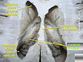
 Coronal section of inferior horn of lateral ventricle.
 -
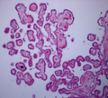 Choroid plexus histology 40x
 -
 Choroid plexus
 -
 Choroid plexus
 -
 Choroid plexus


See also
References
![]() This article incorporates text in the public domain from page 798 of page 841 of page 816 of the 20th edition of Gray's Anatomy (1918)
This article incorporates text in the public domain from page 798 of page 841 of page 816 of the 20th edition of Gray's Anatomy (1918)
- ISBN 978-0-7817-9069-7.
- ^ S2CID 11473603.
- ^ PMID 26174708.
- PMID 22118931.
- PMID 30844183.
- PMID 25837384.
- PMID 29437557.
- ISBN 978-1-4160-4574-8.
- ISBN 978-0-7216-0240-0.
- ^ PMID 29195051.
- PMID 29428972.
- PMID 12553165.
- PMID 17953660.
- ^ a b c d e Laterra J, Keep R, Betz LA, et al. (1999). "Blood–cerebrospinal fluid barrier". Basic Neurochemistry: Molecular, Cellular and Medical Aspects (6th ed.). Philadelphia: Lippincott-Raven.
- PMID 26998242.
The embryonic CSF-brain barrier, shown in Figure 1(f). In the ventricular zone is a temporary barrier between the CSF and brain parenchyma. In early brain development, strap junctions are present between adjacent neuroepithelial cells; these form a physical barrier restricting the movement of larger molecules, such as proteins, but not smaller molecules. At later stages of development and in the adult brain, these strap junctions are no longer present when this interface becomes ependyma.
- S2CID 22154007.
- PMID 24357543.
- ^ PMID 10607945.
- S2CID 40130437.
- PMID 9678699.
- ^ Suzuki, S., Katsumata, T., Ura, R. Fujita, T., Niizima, M. & Suzuki, H. (1936). Über die Nomina Anatomica Nova. Folia Anatomica Japonica, 14, 507-536.
- ^ a b Liddell HG, Scott R (1940). A Greek-English Lexicon. Oxford: Clarendon Press.
Sources
- Brodbelt A, Stoodley M (October 2007). "CSF pathways: a review". British Journal of Neurosurgery. 21 (5): 510–20. S2CID 6901013.
- Strazielle N, Ghersi-Egea JF (July 2000). "Choroid plexus in the central nervous system: biology and physiopathology". Journal of Neuropathology and Experimental Neurology. 59 (7): 561–74. PMID 10901227.
External links
- 3-Dimensional images of choroid plexus (marked red)
- "Anatomy diagram: 13048.000-3". Roche Lexicon - illustrated navigator. Elsevier. Archived from the original on 2012-07-22.
- MedPix Images of Choroid Plexus
- More info at BrainInfo