
Bone tumor
| Bone tumor | |
|---|---|
biopsy[1] | |
| Prognosis | Varies with type[4] |
| Frequency | Common[4] |
A bone tumor is an
Diagnosis is generally by
The most common bone tumor is a
Classification

Bone tumors are traditionally classified as
Bone tumors may be classified as "
Primary bone tumors
Primary tumors of bone can be divided into
Malignant primary bone tumors, known as
Secondary bone tumors
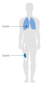
Secondary bone tumors are metastatic lesions which have spread from other organs, most commonly carcinomas of the breast, lung, and prostate. Rarely, primary bone malignancies such as osteosarcoma may also spread to other bones.[12] Reliable and valid statistics on the incidence, prevalence, and mortality of malignant bone tumours are difficult to come by, particularly in older adults (those over 75 years of age) - because
Signs and symptoms
Clinical features of a bone tumor depend on the type of tumor and which part of which bone is affected.[2][13] Symptoms and signs usually result from the pressure effect of the tumor.[1]
There may be a lump, with or without pain.
Diagnosis
A bone tumour may be felt on examination, following which a
Other tests that might be requested include aStaging
-
 Stage 1A bone cancer
 -
 Stage 1B bone cancer
 -
 Stage 2A bone cancer
 -
 Stage 2B bone cancer
 -
 Stage 3 bone cancer


Treatment
Treatment of bone tumors is dependent on the type of tumor.[2] Where available, people with bone tumors are treated at a specialist centre which have surgeons, radiologists, pathologists, oncologists and other support staff.[1] Generally, noncancerous bone tumors may be observed for changes and surgery offered if there is pain or pressure effects on neighbouring body parts. Surgical resection with or without cytotoxic drugs may be considered.[1]
Chemotherapy and radiotherapy
Medication
One of the major concerns is bone density and bone loss. Non-hormonal bisphosphonates increase bone strength and are available as once-a-week prescription pills. Strontium-89 chloride is an intravenous medication given to help with the pain and can be given in three-month intervals.
Surgical treatment
Treatment for some bone cancers may involve
There are other joint preservation surgical reconstruction options, including allograft, tumor-devitalized autograft, vascularized fibula graft, distraction osteogenesis, and custom-made implants.[18] An analysis of massive knee replacements after resection of primary bone tumours showed patients did not score as highly on the Musculoskeletal Tumour Society Score and Knee Society Score as patients who had undergone intra-articular resection.[19]
Thermal ablation techniques
Over the past two decades, CT guided radiofrequency ablation has emerged as a less invasive alternative to surgical resection in the care of benign bone tumors, most notably osteoid osteomas. In this technique, which can be performed under conscious sedation, a RF probe is introduced into the tumor nidus through a cannulated needle under CT guidance and heat is applied locally to destroy tumor cells. Since the procedure was first introduced for the treatment of osteoid osteomas in the early 1990s,[20] it has been shown in numerous studies to be less invasive and expensive, to result in less bone destruction and to have equivalent safety and efficacy to surgical techniques, with 66 to 96% of patients reporting freedom from symptoms.[21][22][23] While initial success rates with RFA are high, symptom recurrence after RFA treatment has been reported, with some studies demonstrating a recurrence rate similar to that of surgical treatment.[24]
Thermal ablation techniques are also increasingly being used in the palliative treatment of painful metastatic bone disease. Currently, external beam radiation therapy is the standard of care for patients with localized bone pain due to metastatic disease. Although the majority of patients experience complete or partial relief of pain following radiation therapy, the effect is not immediate and has been shown in some studies to be transient in more than half of patients.[25] For patients who are not eligible or do not respond to traditional therapies ( i.e. radiation therapy, chemotherapy, palliative surgery, bisphosphonates or analgesic medications), thermal ablation techniques have been explored as alternatives for pain reduction. Several multi-center clinical trials studying the efficacy of RFA in the treatment of moderate to severe pain in patients with metastatic bone disease have shown significant decreases in patient reported pain after treatment.[26][27] These studies are limited however to patients with one or two metastatic sites; pain from multiple tumors can be difficult to localize for directed therapy. More recently, cryoablation has also been explored as a potentially effective alternative as the area of destruction created by this technique can be monitored more effectively by CT than RFA, a potential advantage when treating tumors adjacent to critical structures.[28]
Prognosis
The outlook depends on the type of tumor. The outcome is expected to be good for people with noncancerous (benign) tumors, although some types of benign tumors may eventually become cancerous (malignant). With malignant bone tumors that have not spread, most patients achieve a cure, but the cure rate depends on the type of cancer, location, size, and other factors.[citation needed]
Epidemiology
Bone tumors that originate from bone are very rare and account for around 0.2% of all tumors.
History
The earliest known bone tumor was an osteosarcoma in a foot bone belonging to a person who died in Swartkrans Cave, South Africa, between 1.6 and 1.8 million years ago.[6]
Other animals
Bones are a common site for tumors in cats and dogs.[29]
References
- ^ ISBN 978-1-4441-2098-1.
- ^ a b c d e f g h i j k "Bone Tumor - Types and Treatments - OrthoInfo - AAOS". www.orthoinfo.org. Retrieved 27 June 2021.
- ^ a b c d e f g "Questions and Answers about Bone Cancer" (PDF). Centers for Disease Control and Prevention. Retrieved 27 June 2021.
- ^ ISBN 978-92-832-4502-5.
- ^ a b "SEER Stat Fact Sheets: Bone and Joint Cancer". NCI. Retrieved 18 June 2014.
- ^ a b Strauss, Mark (28 July 2016). "Earliest Human Cancer Found in 1.7-Million-Year-Old Bone". Culture. Archived from the original on March 10, 2021. Retrieved 27 June 2021.
- ^ S2CID 231679037.
- S2CID 231595171.
- ^ Jeon DG, Song WS, Kong CB, Kim JR, Lee SY. MFH of Bone and Osteosarcoma Show Similar Survival and Chemosensitivity. Clin Orthop Rel Res 469;584-90.
- ^ "Multiple Myeloma". The Lecturio Medical Concept Library. Retrieved 26 August 2021.
- PMID 32485873.
- ^ "Osteosarcoma". The Lecturio Medical Concept Library. Retrieved 26 August 2021.
- ISBN 978-1-4471-6577-4.
- PMID 23255735.
- Mount Sinai Hospital, New York
- ^ 10 year survival in Pediatric Osteosarcoma[permanent dead link]
- ^ Survival in Adult Osteosarcoma and MFH of Bone[permanent dead link]
- S2CID 208650189.
- PMID 11021451.
- PMID 1549690.[permanent dead link]
- S2CID 21047698.
- PMID 12944597.[permanent dead link]
- S2CID 21496405.
- S2CID 10709128. Archived from the originalon 2016-10-06. Retrieved 2016-08-07.
- PMID 6178497.
- PMID 20041484.
- PMID 14722039.
- PMID 23065947.
- S2CID 232409416.