
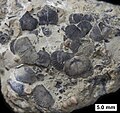
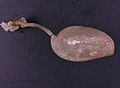
Brachiopod
The article's lead section may need to be rewritten. The reason given is: The current lead near-requires its own sections; not a good summary. (December 2023) |
| Brachiopod Temporal range:
| |
|---|---|

| |
linguliform brachiopod
| |
rhynchonelliform ) brachiopod
| |
| Scientific classification | |
| Domain: | Eukaryota |
| Kingdom: | Animalia |
| Superphylum: | Lophotrochozoa |
| Clade: | Lophophorata |
| Clade: | Brachiozoa |
| Phylum: | Brachiopoda Duméril, 1806[1] |
| Subphyla and classes | |
|
See taxonomy | |
| Diversity[2] | |
| About 100 living genera About 5,000 fossil genera | |
Brachiopods (
Brachiopod lifespans range from three to over thirty years. Ripe gametes (ova or sperm) float from the gonads into the main coelom and then exit into the mantle cavity. The larvae of inarticulate brachiopods are miniature adults, with lophophores that enable the larvae to feed and swim for months until the animals become heavy enough to settle to the seabed. The planktonic larvae of articulate species do not resemble the adults, but rather look like blobs with yolk sacs, and remain among the plankton for only a few days before leaving the water column upon metamorphosing.
While traditional classification of brachiopods separate them into distinct inarticulate and articulate groups, two approaches appeared in the 1990s. One approach groups the inarticulate
It was suggested in 2003 that brachiopods had evolved from an ancestor similar to
Brachiopods live only in the sea, and most species avoid locations with strong currents or waves. The larvae of articulate species settle in quickly and form dense populations in well-defined areas while the larvae of inarticulate species swim for up to a month and have wide ranges. Brachiopods now live mainly in cold water and low light. Fish and crustaceans seem to find brachiopod flesh distasteful and seldom attack them. Among brachiopods, only the lingulids (Lingula sp.[4]) have been fished commercially, on a very small scale. One brachiopod species (Coptothyrus adamsi) may be a measure of environmental conditions around an oil terminal being built in Russia on the shore of the Sea of Japan. The word "brachiopod" is formed from the Ancient Greek words brachion ("arm") and podos ("foot").[5] They are often known as "lamp shells", since the curved shells of the class Terebratulida resemble pottery oil-lamps.[2]
Brachiopods are the state fossil of the U.S. state of Kentucky.[6]
Anatomy
Shell structure and function
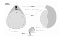
_01_svg.svg)
Pedicle (ventral) valve
Brachial (dorsal) valve
Pedicle
Surface
Modern brachiopods range from 1 to 100 millimetres (0.039 to 3.937 in) long, and most species are about 10 to 30 millimetres (0.39 to 1.18 in).
The brachial valve is usually smaller and bears brachia ("arms") on its inner surface. These brachia are the origin of the phylum's name, and support the lophophore, used for feeding and respiration. The pedicle valve is usually larger, and near the hinge it has an opening for the stalk-like pedicle through which most brachiopods attach themselves to the substrate. (R. C. Moore, 1952) The brachial and pedicle valves are often called the dorsal and ventral valves, respectively, but some paleontologists regard the terms "dorsal" and "ventral" as irrelevant since they believe that the "ventral" valve was formed by a folding of the upper surface under the body. The ventral ("lower") valve actually lies above the dorsal ("upper") valve when most brachiopods are oriented in life position. In many living articulate brachiopod species, both valves are convex, the surfaces often bearing growth lines and/or other ornamentation. However, inarticulate lingulids, which burrow into the seabed, have valves that are smoother, flatter and of similar size and shape. (R. C. Moore, 1952)
Articulate ("jointed") brachiopods have a tooth and socket arrangement by which the pedicle and brachial valves hinge, locking the valves against lateral displacement. Inarticulate brachiopods have no matching teeth and sockets; their valves are held together only by muscles. (R. C. Moore, 1952)
All brachiopods have adductor muscles that are set on the inside of the pedicle valve and which close the valves by pulling on the part of the brachial valve ahead of the hinge. These muscles have both "quick" fibers that close the valves in emergencies and "catch" fibers that are slower but can keep the valves closed for long periods. Articulate brachiopods open the valves by means of abductor muscles, also known as diductors, which lie further to the rear and pull on the part of the brachial valve behind the hinge. Inarticulate brachiopods use a different opening mechanism, in which muscles reduce the length of the coelom (main body cavity) and make it bulge outwards, pushing the valves apart. Both classes open the valves to an angle of about 10 degrees. The more complex set of muscles employed by inarticulate brachiopods can also operate the valves as scissors, a mechanism that lingulids use to burrow.[10]

Each valve consists of three layers, an outer periostracum made of organic compounds and two biomineralized layers. Articulate brachiopods have an outermost periostracum made of proteins, a "primary layer" of calcite (a form of calcium carbonate) under that, and innermost a mixture of proteins and calcite.[10] Inarticulate brachiopod shells have a similar sequence of layers, but their composition is different from that of articulated brachiopods and also varies among the classes of inarticulate brachiopods. The Terebratulida are an example of brachiopods with a punctate shell structure; the mineralized layers are perforated by tiny open canals of living tissue, extensions of the mantle called caeca, which almost reach the outside of the primary layer. These shells can contain half of the animal's living tissue. Impunctate shells are solid without any tissue inside them. Pseudopunctate shells have tubercles formed from deformations unfurling along calcite rods. They are only known from fossil forms, and were originally mistaken for calcified punctate structures.[11][12]
Lingulids and discinids, which have pedicles, have a matrix of glycosaminoglycans (long, unbranched polysaccharides), in which other materials are embedded: chitin in the periostracum;[10] apatite containing calcium phosphate in the primary biomineralized layer;[13] and a complex mixture in the innermost layer, containing collagen and other proteins, chitinophosphate and apatite.[10][14] Craniids, which have no pedicle and cement themselves directly to hard surfaces, have a periostracum of chitin and mineralized layers of calcite.[10][15] Shell growth can be described as holoperipheral, mixoperipheral, or hemiperipheral. In holoperipheral growth, distinctive of craniids, new material is added at an equal rate all around the margin. In mixoperipheral growth, found in many living and extinct articulates, new material is added to the posterior region of the shell with an anterior trend, growing towards the other shell. Hemiperipheral growth, found in lingulids, is similar to mixoperipheral growth but occurs in mostly a flat plate with the shell growing forwards and outwards.[16]
Mantle
Brachiopods, as with
Relatively new cells in a groove on the edges of the mantle secrete material that extends the periostracum. These cells are gradually displaced to the underside of the mantle by more recent cells in the groove, and switch to secreting the mineralized material of the shell valves. In other words, on the edge of the valve the periostracum is extended first, and then reinforced by extension of the mineralized layers under the periostracum.
In most brachiopods, diverticula (hollow extensions) of the mantle penetrate through the mineralized layers of the valves into the periostraca. The function of these diverticula is uncertain and it is suggested that they may be storage chambers for chemicals such as glycogen, may secrete repellents to deter organisms that stick to the shell or may help in respiration.[10] Experiments show that a brachiopod's oxygen consumption drops if petroleum jelly is smeared on the shell, clogging the diverticula.[17]
Lophophore
Like
The tentacles bear
Pedicle and other attachments

Most modern species attach to hard surfaces by means of a cylindrical pedicle ("stalk"), an extension of the body wall. This has a chitinous cuticle (non-cellular "skin") and protrudes through an opening in the hinge.[10] However, some genera have no pedicle, such as the inarticulate Crania and the articulate Lacazella; they cement the rear of the "pedicle" (ventral) valve to a surface so that the front is slightly inclined up away from the surface.[2][10] In these brachiopods, the ventral valve lacks a pedicle opening.[24] In a few articulate genera such as Neothyris and Anakinetica, the pedicles wither as the adults grow and finally lie loosely on the surface. In these genera the shells are thickened and shaped so that the opening of the gaping valves is kept free of the sediment.[2]
Pedicles of inarticulate species are extensions of the main coelom, which houses the internal organs. A layer of longitudinal muscles lines the
The pedicle of articulate brachiopods has no coelom, and its homology is unclear. It is constructed from a different part of the larval body, and has a compact core composed of connective tissue. Muscles at the rear of the body can straighten, bend or even rotate the pedicle. The far end of the pedicle generally has rootlike extensions or short papillae ("bumps"), which attach to hard surfaces. However, articulate brachiopods of the genus Chlidonophora use a branched pedicle to anchor in sediment. The pedicle emerges from the pedicle valve, either through a notch in the hinge or, in species where the pedicle valve is longer than the brachial, from a hole where the pedicle valve doubles back to touch the brachial valve. Some species stand with the front end upwards, while others lie horizontal with the pedicle valve uppermost.[10]
Some early brachiopods—for example strophomenates, kutorginates and obolellates—do not attach using their pedicle, but with an entirely different structure known as the "pedicle sheath", which has no relationship to the pedicle.[25][26] This structure arises from the umbo of the pedicle valve, at the centre of the earliest (metamorphic) shell at the location of the protegulum. It is sometimes associated with a fringing plate, the colleplax.[26]
Biology
Feeding and excretion


The water flow enters the lophophore from the sides of the open valves and exits at the front of the animal. In lingulids the entrance and exit channels are formed by groups of chaetae that function as funnels.
Nutrients are transported throughout the coelom, including the mantle lobes, by cilia.
The majority of food consumed by brachiopods is digestible, with very little solid waste produced.[29] The cilia of the lophophore can change direction to eject isolated particles of indigestible matter. If the animal encounters larger lumps of undesired matter, the cilia lining the entry channels pause and the tentacles in contact with the lumps move apart to form large gaps and then slowly use their cilia to dump the lumps onto the lining of the mantle. This has its own cilia, which wash the lumps out through the opening between the valves. If the lophophore is clogged, the adductors snap the valves sharply, which creates a "sneeze" that clears the obstructions.[17] In some inarticulate brachiopods the digestive tract is U-shaped and ends with an anus that eliminates solids from the front of the body wall.[27] Other inarticulate brachiopods and all articulate brachiopods have a curved gut that ends blindly, with no anus.[10] These animals bundle solid waste with mucus and periodically "sneeze" it out, using sharp contractions of the gut muscles.[17]
Circulation and respiration
The lophophore and mantle are the only surfaces that absorb oxygen and eliminate carbon dioxide. Oxygen seems to be distributed by the fluid of the coelom, which is circulated through the mantle and driven either by contractions of the lining of the coelom or by beating of its cilia. In some species oxygen is partly carried by the respiratory pigment hemerythrin, which is transported in coelomocyte cells.[10] The maximum oxygen consumption of brachiopods is low, and their minimum requirement is not measurable.
Brachiopods also have colorless blood, circulated by a muscular heart lying in the dorsal part of the body above the stomach.[10] The blood passes through vessels that extend to the front and back of the body, and branch to organs including the lophophore at the front and the gut, muscles, gonads and nephridia at the rear. The blood circulation seems not to be completely closed, and the coelomic fluid and blood must mix to a degree.[17] The main function of the blood may be to deliver nutrients.[10]
Nervous system and senses
The "brain" of adult articulates consists of two
Reproduction and life cycle
Lifespans range from 3 to over 30 years.
The
The
Taxonomy
Taxonomic history
Brachiopod fossils show great diversity in the morphology of the shells and lophophore, while the modern genera show less diversity but provide soft-bodied characteristics. Both fossils and extant species have limitations that make it difficult to produce a comprehensive classification of brachiopods based on morphology. The phylum also has experienced significant convergent evolution and reversals (in which a more recent group seems to have lost a characteristic that is seen in an intermediate group, reverting to a characteristic last seen in an older group). Hence some brachiopod taxonomists believe it is premature to define higher levels of classification such as order, and recommend instead a bottom-up approach that identifies genera and then groups these into intermediate groups.[33]
However, other taxonomists believe that some patterns of characteristics are sufficiently stable to make higher-level classifications worthwhile, although there are different views about what the higher-level classifications should be.[33] The "traditional" classification was defined in 1869; two further approaches were established in the 1990s:[14][34]
- In the "traditional" classification, brachiopods are divided into the Articulata and Inarticulata. The Articulata have toothed hinges between the valves, while the hinges of the Inarticulata are held together only by muscles.[10][14]
- A classification devised in the 1990s, based on the materials of which the shells are based, united the
- A three-part scheme, also from the 1990s, places the Craniiformea. The Lingulida and Discinida are grouped as Linguliformea,[35] and the Rhynchonellida and Terebratulida as Rhynchonelliformea.[36][37]
| "Traditional" classification[10][14] | Inarticulata | Articulata | |||
|---|---|---|---|---|---|
| "Calciata" approach[14] | Lingulata | Calciata | |||
| Three-part approach[36][37] | Linguliformea | Craniiformea |
Rhynchonelliformea | ||
| Orders | Lingulida[10] | Discinida[10] | Craniida[10] |
Terebratulida[10] | Rhynchonellida[10] |
| Hinge | Teeth and sockets | ||||
| Anus | On front of body, at end of U-shaped gut | At the back of body | None | ||
| Pedicle | Contains coelom with muscles running through | No pedicle | No coelom, muscles where joins body | ||
| Long, burrows | Short, attached to hard surfaces | None, cemented to surface | Short, attached to hard surfaces[14] | ||
| Periostracum | Glycosaminoglycans and chitin |
Chitin | Proteins | ||
| Primary (middle) mineralized layer of shell | Glycosaminoglycans and apatite (calcium phosphate) | Calcite (a form of calcium carbonate) | |||
| Inner mineralized layer of shell | Collagen and other proteins, chitinophosphate and apatite (calcium phosphate) | Calcite | Proteins and calcite | ||
| Chaetae around opening of shells | Yes[14] | Yes[38] | Yes[14] | ||
| Coelom fully divided | Yes[14] | No[14] | Yes[14] | ||
| Larvae | Planktotrophic (feeding) | Lecithotrophic (non-feeding) | |||
About 330 living species are recognized,[14] grouped into over 100 genera. The great majority of modern brachiopods are rhynchonelliforms (Articulata).[2]
Modern classification
Genetic analysis performed since the 1990s has extended the understanding of the relationship between different organisms. It is now clear the brachiopods do not belong to the
Orders
This shows the taxonomy of brachiopods down to the order level, including extinct groups, which make up the majority of species. Extinct groups are indicated with a (†) symbol:
- Subphylum Linguliformea
- Class Lingulata
- Order Lingulida
- Order †Acrotretida
- Order †Siphonotretida
- Class †Paterinata
- Order †Paterinida
- Order †
- Class Lingulata
- Subphylum Craniiformea
- Class Craniata
- Order Craniida
- Order †Craniopsida
- Order †Trimerellida
- Class Craniata
- Subphylum Rhynchonelliformea
- Class †Chileata
- Order †Chileida
- Order †Dictyonellida
- Order †
- Class †Obolellata
- Order †Obolellida
- Order †Naukatida
- Order †
- Class †Kutorginata
- Order †Kutorginida
- Order †
- Class †Strophomenata
- Order †Billingsellida
- Order †Strophomenida
- Order †Productida
- Order †Orthotetida
- Class Rhynchonellata
- Order Rhynchonellida
- Order Terebratulida
- Order Thecideida
- Order †Protorthida
- Order †Orthida
- Order †Pentamerida
- Order †Atrypida
- Order †Athyridida
- Order †Spiriferida
- Order †Spiriferinida
- Class †Chileata
Ecology

Distribution and habitat
Brachiopods are an entirely marine phylum, with no known freshwater species. Most species avoid locations with strong currents or waves, and typical sites include rocky overhangs, crevices and caves, steep slopes of
Interactions with other organisms
Brachiopods have a low
Brachiopod shells occasionally show evidence of damage by predators, and sometimes of subsequent repair. Fish and crustaceans seem to find brachiopod flesh distasteful.
Among brachiopods only the lingulids have been fished commercially, and only on a very small scale.[44] It is mostly the fleshy pedicle that is eaten.[45][46][47][48] Brachiopods seldom settle on artificial surfaces, probably because they are vulnerable to pollution. This may make the population of Coptothyrus adamsi useful as a measure of environmental conditions around an oil terminal being built in Russia on the shore of the Sea of Japan.[1]
Evolutionary history

Fossil record
Over 12,000 fossil species are recognized,
Since 1991 Claus Nielsen has proposed a hypothesis about the development of brachiopods, adapted in 2003 by Cohen and colleagues as a hypothesis about the earliest evolution of brachiopods. This "brachiopod fold" hypothesis suggests that brachiopods evolved from an ancestor similar to
However, fossils from 2007 onwards have supported a new interpretation of the Early-Cambrian tommotiids, and a new hypothesis that brachiopods evolved from tommotiids. The "armor mail" of tommotiids was well-known but not in an assembled form, and it was generally assumed that tommotiids were slug-like animals similar to Halkieria, except that tommotiids' armor was made of organophosphatic compounds while that of Halkieria was made of calcite. However, fossils of a new tommotiid, Eccentrotheca, showed an assembled mail coat that formed a tube, which would indicate a sessile animal rather than a creeping slug-like one. Eccentrotheca's organophosphatic tube resembled that of phoronids,[54] sessile animals that feed by lophophores and are regarded either very close relatives or a sub-group of brachiopods.[55] Paterimitra, another mostly assembled fossil found in 2008 and described in 2009, had two symmetrical plates at the bottom, like brachiopod valves but not fully enclosing the animal's body.[56]
At their peak in the
Brachiopod fossils have been useful indicators of climate changes during the Paleozoic era. When global temperatures were low, as in much of the Ordovician, the large difference in temperature between equator and poles created different collections of fossils at different latitudes. On the other hand, warmer periods, such much of the Silurian, created smaller difference in temperatures, and all seas at the low to middle latitudes were colonized by the same few brachiopod species.[61]
Evolutionary family tree

Deuterostomes or protostomes
From about the 1940s to the 1990s,
- Radial cleavage in the earliest divisions of the egg appears to be the original condition for the ancestral bilaterians, in the earliest Ecdysozoa and possibly in the earliest Eutrochozoa, a major sub-group of the Lophotrochozoa.[66] Hence radial cleavage does not imply that brachiopods are affiliated with deuterostomes.[65]
- The traditional view is that the coelom(s) in deuterostomes and protostomes form by different process, called enterocoely and schizocoely, respectively.[65] However, research since the early 1990s has found significant exceptions.[66][67] Both types of coelom construction appear among brachiopods, and therefore do not imply that brachiopods are deuterostomes.[65]
- The terms "deuterostomes" and "protostomes" originally defined distinct ways of forming the mouth from the blastopore, a depression that appears in an early stage of the embryo. However, some "protostomes" form the mouth using a process more like that typical of deuterostomes.[68][69] Hence forming the mouth via a deuterostome-like process does not imply that brachiopods are affiliated with deuterostomes.[65]
Nielsen views the brachiopods and closely related
From 1988 onwards analyses based on
Some combined studies in 2000 and 2001, using both molecular and morphological data, support brachiopods as Lophotrochozoa,[72][73] while others in 1998 and 2004 concluded that brachiopods were deuterostomes.[71]
Relationship with other lophotrochozoans
The phoronids feed with a lophophore, burrow or encrust on surfaces, and build three-layered tubes made of polysaccharide, possibly chitin, mixed with particles with seabed material. Traditionally they have been regarded as a separate phylum, but increasingly detailed molecular phylogeny studies between 1997 and 2000 have concluded that phoronids are a sub-group of brachiopods.[55] However, an analysis in 2005 concluded that phoronids are a sub-group of bryozoans.[74]
While all molecular phylogeny studies and half the combined studies until 2008 conclude that brachiopods are
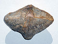
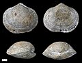
Gallery
-
 Brachiopod morphology
 -
 Cranaena, a terebratulid from the Middle Devonian of Wisconsin.
 -
 The Carboniferous brachiopod Neospirifer condor from Bolivia. The specimen is 7 cm across.
 -
 Tylothyris, a spiriferid from the Middle Devonian of Wisconsin
 -
 Rhynchotrema dentatum, a rhynchonellid brachiopod from the Cincinnatian (Upper Ordovician) of southeastern Indiana
 -
 A Devonian spiriferid brachiopod from Ohio that served as a host substrate for a colony of hederellids. The specimen is 5 cm wide.
 -
 Syringothyris texta (Hall 1857), dorsal view, internal mold. Lower Carboniferous of Wooster, Ohio
 -
 Petrocrania brachiopods attached to a strophomenid brachiopod; Upper Ordovician of southeastern Indiana.
 -
 Ozamis City, Philippines
 -
 Barroisella, a lingulid from the Middle Devonian of Wisconsin.
 -
 Brachiopod casts in the Lock Haven Formation
 -
 Hercosestria cribrosa Cooper & Grant 1969 (Roadian, Guadalupian, Middle Permian); Glass Mountains, Texas.
 -
 Productid brachiopod ventral valve interior; Roadian, Guadalupian (Middle Permian); Glass Mountains, Texas.
 -
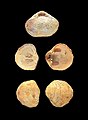 Terebratella sanguinea (Leach, 1814)
 -
 Schizophoria, an orthid from the Middle Devonian of Wisconsin.
 -
 Striatochonetes, a chonetid from the Middle Devonian of Wisconsin.
 -
 Oleneothyris, a terebratulid from the Cenozoic of New Jersey
 -
 AMagellania joubini from the Ross Sea
 -
 A mass burial of brachiopods from the Logan Formation (Mississippian) in Wooster, Ohio
 -
 An Abyssothyris from the Challenger Plateau in the Pacific
 -


_(Tertiary;_New_Egypt,_New_Jersery,_USA)_7.jpg)


.jpg)
_(modern;_offshore_California,_USA)_4.jpg)
See also
- Taxonomy of the Brachiopoda
- Evolution of brachiopods
- List of brachiopod genera
- List of brachiopod species
- Novocrania anomala
- Margaret Jope
Notes
- ^ a b Zvyagintsev etc: Brachio fouling & (2007).
- ^ a b c d e f g h i j k l m n o Cohen: Brachiopoda ELS & (2002).
- doi:10.18268/BSGM2016v68n2a9.)
{{cite journal}}: CS1 maint: multiple names: authors list (link - S2CID 129829675.
- ^ Shorter Oxford English Dictionary & (2002), entry "Brachiopod".
- ^ "Kentucky's State Fossil: Brachiopods". Kentucky Geological Survey.
- PMID 29690189.
- ISBN 978-0-8137-3015-8.
- PMID 36123753.
- ^ a b c d e f g h i j k l m n o p q r s t u v w x y z aa ab ac ad ae af ag ah ai aj ak al am an ao ap aq Ruppert etc: Invert Zoo & (2004), pp. 821–829, ch. "Lophophorata" sect. "Brachiopoda".
- ^ Marine Species Identification Portal : Brachiopoda of the North Sea
- ISBN 978-1-351-46309-6.
- ^ "Apatite" is strictly defined in terms of its structure rather than chemical composition. Some forms contain calcium phosphate and others have calcium carbonate. See Cordua, W.S. "Apatite Ca5(PO4, CO3)3(F, Cl, OH) Hexagonal". University of Wisconsin. Archived from the original on 30 August 2009. Retrieved 23 October 2009.
- ^ a b c d e f g h i j k l m n o Ax: Multicellular Animals & (2003), pp. 87–93, ch."Brachiopoda".
- ^ Parkinson etc: Brachiopod shells & (2005).
- ISBN 9780922152766.
- ^ a b c d e f g h i j k l m n Doherty: Lophophorates & (2001), pp. 341–342, 356–363, sect. "Introduction", "Brachiopoda".
- ^ Ruppert etc: Invert Zoo & (2004), pp. 829–845, ch. "Lophophorata" sect. "Bryozoa".
- ^ Ruppert etc: Invert Zoo & (2004), pp. 817–821, ch. "Lophophorata" sect. "Phoronida".
- PMID 34376709.
- ^ Ruppert etc: Invert Zoo & (2004), p. 817, ch. "Lophophorata" sect. "Introduction".
- ^ Riisgård etc: Downstream & (2000).
- ^ a b Emig: Inart Brach & (2001).
- ^ Wells, Roger M. "BRACHIOPODA MORPHOLOGY AND ECOLOGY". Invertebrate Paleontology Tutorial. State University of New York College at Courtland. Archived from the original on 20 June 2010. Retrieved 6 March 2010.
- S2CID 134399842.
- ^ doi:10.1111/let.12204.)
{{cite journal}}: CS1 maint: multiple names: authors list (link - ^ a b c d Cohen etc: Brachiopod fold & (2003).
- ^ Ruppert etc: Invert Zoo & (2004), pp. 212–214, ch. "Bilateria" sect. "Excretion".
- ^ Cowen: History of life & (2000), p. 408, ch. "Invert Paleo".
- ^ Nielsen: Brachio brains & (2005).
- ^ Ruppert etc: Invert Zoo & (2004), pp. 214–219, ch. "Bilateria" sect. "Reproduction".
- PMID 28879180.
- ^ a b Carlson: Ghosts & (2001).
- ^ ITIS: Brachiopoda.
- doi:10.18268/BSGM2016v68n2a9.)
{{cite journal}}: CS1 maint: multiple names: authors list (link - ^ a b Milsom etc: 3-part taxonomy & (2009).
- ^ a b Williams etc: Suprafamilial Classif & (2000), pp. xxxix–xlv, Preface.
- ^ "Marginal mantle setae were long thought to be absent in craniides, but NIELSEN (1991) has now demonstrated their presence in juvenile Neocrania." -- Williams, Alwyn; Brunton, C.H.C.; Carlson, S.J.; et al. (1997–2007). Kaesler, Roger L.; Selden, Paul (eds.). Part H, Brachiopoda (Revised). Treatise on Invertebrate Paleontology. Boulder, Colorado; Lawrence, Kansas: Geological Society of America; University of Kansas.
- PMID 10714876.
- .
- ^ Vermeij: Directionality & (1999).
- ^ Kowalewski etc: 2nd-choice prey & (2002).
- ^ Iyengar: Kleptoparasitism & (2008).
- ^ UCMP: Lingulata.
- ^ The world’s oldest food delicacy revealed
- ^ The Potency and Food Safety of Lamp Shells (Brachiopoda: Lingula sp.) as Food Resources
- ^ Applied Palaeontology
- ^ Fishing in Many Waters
- ^ a b UCMP: Brachio Fossil Range.
- ^ a b Fortey: Fossils the key & (2008), ch. "How to recognize" sect. "Brachiopods".
- ^ Ushatinskaya: Earliest brachiopods & (2008).
- ^ Balthasar etc: Brachios stem Phoronids & (2009).
- ^ Conway Morris etc: Articulated Halkieriids & (1995).
- .
- ^ a b c Cohen: Phoronids in Brachios, (2000 & ).
- PMID 19203919.
- ISBN 9780262354189. Retrieved 2022-08-23.
- ^ a b c Barry: Great Dying & (2002).
- ^ Gould etc: Clams and Brachios & (1980).
- ^ Knoll etc: P-Tr extinction & (2007).
- ^ Gaines etc: Inverteb proxies & (2009).
- ^ a b Halanych: New phylogeny & (2004).
- ^ De Rosa (2001) cites the following examples of brachiopods as close to deuterostomes:
- Hyman, L.H. (1940). The invertebrates. McGraw-Hill. ISBN 978-0-07-031661-4.
- Eernisse, D.J.; Albert, J.S.; Anderson, F.E. (1992). "Annelida and Arthropoda are not sister taxa: A phylogenetic analysis of spiralian metazoan morphology". Systematic Biology. 41 (3): 305–330. .
- Nielsen, C. (1995). Animal evolution: Interrelationships of the living phyla. Oxford University Press. ISBN 978-0-19-854867-6.
- Lüter, C.; Bartholomaeus, T. (July 1997). "The phylogenetic position of Brachiopoda—a comparison of morphological and molecular data". Zoologica Scripta. 26 (3): 245–253. S2CID 83934233.
- Hyman, L.H. (1940). The invertebrates. McGraw-Hill.
- ^ UCMP: Deuterostomia.
- ^ PMID 12116636.
- ^ a b Valentine: Cleavage patterns & (1997).
- ^
- Ruppert, E.E (1991). "Introduction to the aschelminth phyla: A consideration of mesoderm, body cavities, and cuticle". In Harrison, F.W.; Ruppert, E.E (eds.). Microscopic anatomy of invertebrates, volume 4: Aschelminthes. Wiley-Liss. pp. 1–17.
- Budd, G.E; Jensen, S (May 2000). "A critical reappraisal of the fossil record of the bilaterian phyla". Biological Reviews of the Cambridge Philosophical Society. 75 (2): 253–295. S2CID 39772232.
- ISBN 978-0-08-017069-5.
- S2CID 14964932.
- ^ Nielsen: Phylog pos of Brachios & (2002).
- ^ a b c d Helmkampf etc: Lophotrochozoa concept & (2008).
- ^ Giribet etc: Combined phylogeny & (2000).
- ^ Peterson etc: Combined phylogeny & (2001).
- ^ Wood etc: Phylactolaemate Phylog & (2005).
- ^ Dunn etc: Close to Nemertines & (2008).
- ^ Bourlat etc: Close to Nemertines & (2008).
References
- R.C.Moore, 1952; Brachiopods in Moore, Lalicher, and Fischer; Invertebrate Fossils, McGraw-Hill.
- Ax, P (2003). Multicellular Animals: Order in Nature - System Made by Man. Multicellular Animals: A New Approach to the Phylogenetic Order in Nature. Vol. 3. Springer. ISBN 978-3-540-00146-1. Retrieved 2 November 2009.
- Barry, P.L (January 28, 2002). "The Great Dying". Science@NASA. Science and Technology Directorate, Marshall Space Flight Center, NASA. Archived from the original on April 7, 2009. Retrieved March 26, 2009.
- Balthasar, U; Butterfield, N.J (2009). "Early Cambrian "soft−shelled" brachiopods as possible stem−group phoronids". Acta Palaeontologica Polonica. 54 (2): 307–314. S2CID 54072910.
- Bourlat, S.J; Nielsen, C.; Economou, A.D.; Telford, M.J (October 2008). "Testing the new animal phylogeny: A phylum level molecular analysis of the animal kingdom". Molecular Phylogenetics and Evolution. 49 (1): 23–31. PMID 18692145.
- Carlson, S.J (November 2001). "Ghosts of the Past, Present, and Future in Brachiopod Systematics". Journal of Paleontology. 75 (6): 1109–1118. S2CID 85822720.
- Cohen, B.L (2000). "Monophyly of brachiopods and phoronids: reconciliation of molecular evidence with Linnaean classification (the subphylum Phoroniformea nov.)" (PDF). Proceedings of the Royal Society B. 267 (1440): 225–231. PMID 10714876. Retrieved 31 Jan 2010.
- Cohen, B.L; Holmer, L. E.; Lüter, C (2003). "The brachiopod fold: a neglected body plan hypothesis" (PDF). Palaeontology. 46 (1): 59–65. . Retrieved 2 November 2009.
- Cohen, B.L (2006). "Brachiopoda". Encyclopedia of Life Sciences. John Wiley & Sons, Ltd. ISBN 978-0470016176.
- Conway Morris, S; Peel, J. S (1995). "Articulated Halkieriids from the Lower Cambrian of North Greenland and their Role in Early Protostome Evolution". Philosophical Transactions of the Royal Society B. 347 (1321): 305–358. .
- Cowen, R (2000). "Invertebrate paleontology". History of life (3 ed.). Wiley-Blackwell. p. 408. ISBN 978-0-632-04444-3. Retrieved 2 November 2009.
- Doherty, P.J (2001). "The Lophophorates". In Anderson, D.T (ed.). Invertebrate Zoology (2 ed.). Oxford University Press. pp. 356–363. ISBN 978-0-19-551368-4.
- Dunn, C.W; Hejnol, A.; Matus, D.Q.; Pang, K.; Browne, W.E.; Smith, S.A.; Seaver, E.; Rouse, G.W.; et al. (2008). "Broad phylogenomic sampling improves resolution of the animal tree of life". Nature. 452 (7188): 745–749. S2CID 4397099.
- Emig, C.C (1997). "Ecology of Inarticulated Brachiopods" (PDF). In Kaesler, R.L (ed.). Treatise on Invertebrate Paleontology. Vol. Part H. Geological Society of America. pp. 473–481. Retrieved 14 November 2009.
- ISBN 978-0-674-31135-0. Retrieved 16 November 2009.
- Gaines, R.R; Droser (2009). "Animal proxies - Invertebrates". In Gornitz, V (ed.). Encyclopedia of paleoclimatology and ancient environments. M.L. Springer. p. 11. ISBN 978-1-4020-4551-6. Retrieved 15 November 2009.
- Giribet, G; Distel, D.L.; Polz, M.; Sterrer, W.; Wheeler W. (Sep 2000). "Triploblastic relationships with emphasis on the acoelomates and the position of Gnathostomulida, Cycliophora, Plathelminthes, and Chaetognatha: a combined approach of 18S rDNA sequences and morphology". Systematic Biology. 49 (3): 539–62. PMID 12116426.
- Gould, S.J; Calloway, C.B (1980). "Clams and Brachiopods - Ships that Pass in the Night". Paleobiology. 6 (4): 383–396. S2CID 132467749.
- Halanych, K.M (2004). "The new view of animal phylogeny" (PDF). Annual Review of Ecology, Evolution, and Systematics. 35: 229–256. . Retrieved 2009-04-17.
- Helmkampf, M; Bruchhaus, I.; Hausdorf, B (August 2008). "Phylogenomic analyses of lophophorates (brachiopods, phoronids and bryozoans) confirm the Lophotrochozoa concept". Proceedings of the Royal Society B. 275 (1645): 1927–1933. PMID 18495619.
- Iyengar, E.V (March 2008). "Kleptoparasitic interactions throughout the animal kingdom and a re-evaluation, based on participant mobility, of the conditions promoting the evolution of kleptoparasitism". Biological Journal of the Linnean Society. 93 (4). Wiley InterScience: 745–762. .
- Knoll, A.H.; Bambach, R.K.; Payne, J.L.; Pruss, S. & Fischer, W.W (2007). "Paleophysiology and end-Permian mass extinction" (PDF). Earth and Planetary Science Letters. 256 (3–4): 295–313. doi:10.1016/j.epsl.2007.02.018. Archived from the original(PDF) on 2020-10-04. Retrieved 2008-07-04.
- Kowalewski, M; Hoffmeister, A.P.; Baumiller, T.K.; Bambach, R.K (June 2005). "Secondary Evolutionary Escalation Between Brachiopods and Enemies of Other Prey". Science. 308 (5729): 1774–1777. S2CID 29970931.
- "ITIS: Brachiopoda". Integrated Taxonomic Information System. Retrieved 16 Nov 2009.
- Milsom, C; ISBN 978-1-4051-9336-8. Retrieved 16 November 2009.
- Nielsen, C (2002). "The Phylogenetic Position of Entoprocta, Ectoprocta, Phoronida, and Brachiopoda". Integrative and Comparative Biology. 42 (3): 685–691. PMID 21708765.
- Nielsen, C (2005). "Larval and adult brains". Evolution & Development. 7 (5). Wiley InterScience: 483–489. S2CID 86791922.
- Parkinson, D; Curry, G.B.; Cusack, M.; Fallick, A.E (2005). "Shell structure, patterns and trends of oxygen and carbon stable isotopes in modern brachiopod shells". Chemical Geology. 219 (1–4): 193–235. .
- Peterson, K.J; Eernisse, D.J (May 2001). "Animal phylogeny and the ancestry of bilaterians: inferences from morphology and 18S rDNA gene sequences". Evolution and Development. 3 (3): 170–205. S2CID 7829548.
- Riisgård, H.U; Nielsen, C.; Larsen, P.S (2000). "Downstream collecting in ciliary suspension feeders: the catch-up principle" (PDF). Marine Ecology Progress Series. 207: 33–51. . Retrieved 12 September 2009.
- de Rosa, R (2001). "Molecular Data Indicate the Protostome Affinity of Brachiopods". Systematic Biology. 50 (6): 848–859. PMID 12116636.
- Ruppert, E.E; Fox, R.S.; Barnes, R.D (2004). Invertebrate Zoology (7 ed.). Brooks / Cole. ISBN 978-0-03-025982-1.
- Skovsted, C. B.; Brock, G. A.; Paterson, J. R.; Holmer, L. E.; Budd, G. E. (2008). "The scleritome of Eccentrotheca from the Lower Cambrian of South Australia: Lophophorate affinities and implications for tommotiid phylogeny". Geology. 36 (2): 171. .
- Trumble, W; Brown, L (2002). Shorter Oxford English Dictionary. Oxford University Press, USA. ISBN 978-0-19-860457-0.
- "Introduction to the Lingulata". University of California Museum of Paleontology. Retrieved 16 Nov 2009.
- "Fossil Range Chart of Brachiopods". University of California Museum of Paleontology. Retrieved 16 Nov 2009.
- "Introduction to the Deuterostomia". University of California Museum of Paleontology. Retrieved 27 January 2010.
- Ushatinskaya, G.T (2008). "Origin and dispersal of the earliest brachiopods". Paleontological Journal. 42 (8). Springer Verlag: 776–791. S2CID 83653102.
- Valentine, J.W (1997). "Cleavage patterns and the topology of the metazoan tree of life". Proceedings of the National Academy of Sciences. 94 (15): 8001–8005. PMID 9223303.
- Vermeij, G.T (March 1999). "Inequality and the Directionality of History". The American Naturalist. 153 (3). The American Society of Naturalists: 243–253. S2CID 4378024.
- Williams, A; Carlson, C.H.C; Brunton, S.J (2000). "Outline of Suprafamilial Classification and Authorship". In Williams, A.; Carlson, C.H.C; Brunton, S.J (eds.). Brachiopoda. Geological Society of America and The University of Kansas. pp. xxxix–xlv. )
- Wood, T.S; Lore (2005). "The higher phylogeny of phylactolaemate bryozoans inferred from 18S ribosomal DNA sequences". In Moyano, H. I.; Cancino, J. M.; Wyse-Jackson, P.N (eds.). Bryozoan Studies 2004: Proceedings of the 13th International Bryozoology Association. M. London: Taylor & Francis Group. pp. 361–367.
- Zvyagintsev, A.Y; Radashevsky, V. I.; Kashin, I. A (August 2007). "First record of a brachiopod (Brachiopoda: Terebrataliidae) in the fouling of hydrotechnical installations in Peter the Great Bay, Sea of Japan". Russian Journal of Marine Biology. 33 (4): 264–266. S2CID 23370813.
Further reading
- Candela, Y. (2003). "Late Ordovician brachiopods of the Bardahessiagh Formation of Pomeroy, Ireland". Monograph of the Palaeontographical Society London (618, part of Volume 156): 1–95, pls 1–12.
- Harper, D.A.T. (2006) [1984]. "Brachiopods from the Upper Ardmillan succession (Ordovician) of the Girvan district, Scotland. Parts 1-3". Monograph of the Palaeontographical Society London: 1–187, pls 1–33.
- Moore, R.C.; Lalicker, C.G.; Fischer, A.G. (June 1952). Invertebrate Fossils. Mcgraw-Hill College. ISBN 978-0-07-043020-4.
- Vinn, O.; Wilson, M.A. & Toom, U. (2014). "Earliest rhynchonelliform brachiopod parasite from the Late Ordovician of northern Estonia (Baltica)". Palaeogeography, Palaeoclimatology, Palaeoecology. 411: 42–45. . Retrieved 2014-01-09.
External links
- The Evolution of Brachiopoda – 2016 overview paper by Sandra J. Carlson, Department of Earth and Planetary Sciences, University of Davis, California, at Annual Reviews Archived 2020-04-23 at the Wayback Machine
- BrachNet
- Brachiopoda Database
- Brachiopoda World Database - used from 1995 to 2015
- Information from the Kansas Geological Survey
- Site of R.Filippi
- UC-Berkeley Museum of Paleontology
- Palaeos Brachiopoda (last updated 2002)
|  | |||||||||||||||||||||||||||