
Brainstem
This article needs additional citations for verification. (January 2013) |
| Brainstem | |
|---|---|
 The three distinct parts of the brainstem are colored in this sagittal section of a human brain. | |
| Details | |
| Part of | Brain |
| Parts | Medulla, Pons, Midbrain |
| Identifiers | |
| Latin | truncus encephali |
| MeSH | D001933 |
| NeuroNames | 2052, 236 |
| NeuroLex ID | birnlex_1565 |
| TA98 | A14.1.03.009 |
| TA2 | 5856 |
| FMA | 79876 |
| Anatomical terms of neuroanatomy | |

The brainstem (or brain stem) is the stalk-like[1]: 152 part of the brain that interconnects the cerebrum and diencephalon with the spinal cord.[2] In the human brain, the brainstem is composed of the midbrain, the pons, and the medulla oblongata.[3][1]: 152 The midbrain is continuous with the thalamus of the diencephalon through the tentorial notch.[1]: 152
The brainstem is very small, making up around only 2.6 percent of the brain's total weight.
Structure
The parts of the brainstem are the midbrain, the pons, and the medulla oblongata; the diencephalon is sometimes considered part of the brainstem.[7]: 248
The brainstem extends from just above the
Midbrain


The
The
The ventral tegmental area (VTA) is composed of paired cerebral peduncles. These transmit axons of upper motor neurons.
Midbrain nuclei
The midbrain consists of:
- gray matter around the cerebral aqueduct contains various neurons involved in the pain desensitization pathway. Neurons synapse here. When stimulated by a signal, the synaptic connections activate neurons in the nucleus raphe magnus; the pathway then projects down into the posterior grey columnof the spinal cord which prevents pain sensation transmission.
- Oculomotor nerve nucleus: This is the third cranial nerve nucleus.
- Trochlear nerve nucleus: This is the fourth cranial nerve.
- Red nucleus: This is a motor nucleus that sends a descending tract to the lower motor neurons.
- Substantia nigra pars compacta: This is a concentration of neurons in the ventral portion of the midbrain that uses dopamine as its neurotransmitter and is involved in both motor function and emotion. Its dysfunction is implicated in Parkinson's disease.
- Reticular formation: This is a large area in the midbrain that is involved in various important functions of the midbrain. In particular, it contains lower motor neurons, is involved in the pain desensitization pathway, is involved in the arousal and consciousness systems, and contains the locus coeruleus, which is involved in intensive alertness modulation and in autonomic reflexes.
- Central tegmental tract: Directly anterior to the floor of the fourth ventricle, this is a pathway by which many tracts project up to the cortex and down to the spinal cord.
- Ventral tegmental area: A dopaminergic nucleus, known as group A10 cells[8] is located close to the midline on the floor of the midbrain.
- Rostromedial tegmental nucleus: A GABAergic nucleus located adjacent to the ventral tegmental area.
-
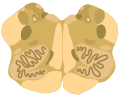
 Cross-section of the midbrain at the level of the superior colliculus
 -
 Cross-section of the midbrain at the level of the inferior colliculus


Pons
This section needs expansion. You can help by adding to it. (April 2014) |
The pons lies between the midbrain and the medulla oblongata. It is separated from the midbrain by the
-
 Cross-section of the middle pons (at the level of cranial nerve V)
 -
 Cross-section of the inferior pons (at the level of the facial genu)


Medulla oblongata
This section needs expansion. You can help by adding to it. (April 2014) |
The
-
 Cross-section of the rostral (superior) medulla
 -
 Cross-section of the middle medulla
 -
 Cross-section of the inferior medulla


Appearance
- From the front

In the medial part of the
The
Between the two pyramids can be seen a decussation of fibers which marks the transition from the medulla to the spinal cord. The medulla is above the decussation and the spinal cord below.
- From behind

The most medial part of the
Superior to the obex is the floor of the fourth ventricle. In the floor of the fourth ventricle, various nuclei can be visualized by the small bumps that they make in the overlying tissue. In the midline and directly superior to the obex is the
Blood supply

The main supply of blood to the brainstem is provided by the basilar arteries and the vertebral arteries.[11]: 740
Development
The human brainstem emerges from two of the three
Function
The brainstem plays important functions in breathing, heart rate, arousal / consciousness, sleep / wake functions and attention / concentration.[12]
There are three main functions of the brainstem:
- The brainstem plays a role in conduction. That is, all information relayed from the body to the cerebrum and cerebellum and vice versa must traverse the brainstem. The ascending pathways coming from the body to the brain are the sensory pathways and include the posterior horn. In addition, there are upper motor neurons that originate in the brainstem's vestibular, red, tectal, and reticular nuclei, which also descend and synapse in the spinal cord.
- The cranial nerves III-XII emerge from the brainstem.[13] These cranial nerves supply the face, head, and viscera. (The first two pairs of cranial nerves arise from the cerebrum).
- The brainstem has integrative functions being involved in cardiovascular system control, respiratory control, pain sensitivity control, alertness, awareness, and consciousness. Thus, brainstem damage is a very serious and often life-threatening problem.
Cranial nerves

This section needs expansion. You can help by adding to it. (October 2016) |
Ten of the twelve pairs of cranial nerves either target or are sourced from the brainstem nuclei.[11]: 725 The nuclei of the oculomotor nerve (III) and trochlear nerve (IV) are located in the midbrain. The nuclei of the trigeminal nerve (V), abducens nerve (VI), facial nerve (VII) and vestibulocochlear nerve (VIII) are located in the pons. The nuclei of the glossopharyngeal nerve (IX), vagus nerve (X), accessory nerve (XI) and hypoglossal nerve (XII) are located in the medulla. The fibers of these cranial nerves exit the brainstem from these nuclei.[14]
Clinical significance
Diseases of the brainstem can result in abnormalities in the function of cranial nerves that may lead to visual disturbances, pupil abnormalities, changes in sensation, muscle weakness, hearing problems, vertigo, swallowing and speech difficulty, voice change, and co-ordination problems. Localizing neurological lesions in the brainstem may be very precise, although it relies on a clear understanding on the functions of brainstem anatomical structures and how to test them.
Brainstem stroke syndrome can cause a range of impairments including locked-in syndrome.
Duret haemorrhages are areas of bleeding in the midbrain and upper pons due to a downward traumatic displacement of the brainstem.[7]: 842
Cysts known as syrinxes can affect the brainstem, in a condition, called syringobulbia. These fluid-filled cavities can be congenital, acquired or the result of a tumor.
Criteria for claiming
Additional images
-
 The midbrain, pons, and medulla oblongata are labelled on this coronal section of the human brain.
 -
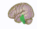 3D visualization of the brainstem in an average human brain
_section_description.JPG)

See also
- Triune brain - reptilian brain
References
- ^ ISBN 9780323396325.
- ISBN 9788131237274.
- ^ ISBN 978-0-7295-3752-0.
- ^ a b c "Brainstem | Definition, Structure, & Function". Encyclopedia Britannica. Retrieved 2020-05-13.
- ^ "Cranial Nerve Nuclei and Brain Stem Circulation". Neuroanatomy Online. Retrieved 2020-05-13.
- ^ Kolb, B. & Whishaw, I. Q. (2009). Fundamentals of human neuropsychology: Sixth edition. New York, NY: Worth Publishers.
- ^ ISBN 978-1-4160-6257-8.
- PMID 23603197.
- ISBN 0683014552.
- PMID 25121239.
- ^ ISBN 978-0-87893-695-3.
- ^ Simplified Brain Behavior Relationships, Alexandria, Virginia, US: Brain Injury Association of Virginia, 2005
- ^ "Lecture 6: Cranial Nerves" (PDF). Archived (PDF) from the original on 2013-04-18. Retrieved 2012-11-10.
- ISBN 978-1-118-49201-7.
- ^ Black's Medical Dictionary 39th edition 1999