
Calcaneus
| Calcaneus | |
|---|---|
 The calcaneus forms the bony part of the heel. It forms a joint with the talus bone, the subtalar joint. | |
 Bones of the foot, with the calcaneus shown in red | |
| Details | |
| Identifiers | |
| Latin | calcaneus, calcaneum, os calcis |
| MeSH | D002111 |
| TA98 | A02.5.11.001 |
| TA2 | 1468 |
| FMA | 24496 |
| Anatomical terms of bone | |
In humans and many other primates, the calcaneus (/kælˈkeɪniəs/; from the Latin calcaneus or calcaneum, meaning heel;[1] pl.: calcanei or calcanea) or heel bone is a bone of the tarsus of the foot which constitutes the heel. In some other animals, it is the point of the hock.
Structure
In humans, the calcaneus is the largest of the
There is a large calcaneal tuberosity located posteriorly on plantar surface with medial and lateral tubercles on its surface. Besides, there is another peroneal tubercle on its lateral surface.
On the lateral side is commonly a tubercle called the calcaneal tubercle (or trochlear process). This is a raised projection located between the tendons of the
Its chief anatomical significance is as a point of divergence of the previously common pathway shared by the distal tendons of peroneus longus and peroneus brevis en route to their distinct respective attachment sites. [3]
The calcaneus is part of two joints: the proximal intertarsal joint and the
Development
In the calcaneus, an ossification center develops during the 4th–7th week of fetal development. [3]
Function
Three muscles insert on the calcaneus: the
 |
 |
| Muscle | Direction | Attachment[4] |
|---|---|---|
| Gastrocnemius | Insertion | Calcaneal tubercle through the achilles tendon |
| Soleus | Insertion | Calcaneal tubercle through the achilles tendon |
| Plantaris | Insertion | Calcaneal tubercle either directly or through the achilles tendon |
| Extensor digitorum brevis | Origin | Dorsal side of calcaneus |
| Abductor hallucis | Origin | Medial process of calcaneus |
| Extensor hallucis brevis | Origin | Dorsal side of calcaneus |
| Abductor digiti minimi | Origin | Calcaneal tubercle |
| Flexor digitorum brevis | Origin | Calcaneal tubercle |
| Quadratus plantae | Origin | Lateral and medial processes of calcaneus |
Clinical significance

Normally the tibia sits vertically above the calcaneus (pes rectus). If the calcaneal axis between these two bones is turned medially the foot is in an everted position (pes valgus), and if it is turned laterally the foot is in an inverted position (pes varus).[5]
- Calcaneal fracture, also known as lover's fracture and Don Juan fracture
Disease
The talar shelf is typically involved in subtalar or talocalcaneal tarsal coalition.
See also
- Calcar
- Bohler's angle
- Sanders classification
Additional images
-
 Left calcaneus. Animation.
 -
 Bones of foot
 -
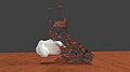 3D rendering of a left calcaneus derived from CT scan data. The calcaneus is white, and the other bones of the foot and ankle are clear to illustrate the position and relationship of the calcaneus to the other tarsal bones.


Notes
- ^ Mosby’s Medical, Nursing and Allied Health Dictionary, Fourth Edition, Mosby-Year Book Inc., 1994, p. 242
- ^ ISBN 9780702029714.
- ^ a b c d Platzer (2004), p 216
- ISBN 978-87-628-0307-7.
- ^ Thieme Atlas of Anatomy (2006), p 410
References
- Platzer, Werner (2004). Color Atlas of Human Anatomy, Vol. 1: Locomotor System (5th ed.). ISBN 3-13-533305-1.
- Thieme Atlas of Anatomy: General Anatomy and Musculoskeletal System. Thieme. 2006. ISBN 1-58890-419-9.
- Saladin, Kenneth (2012). Anatomy and Physiology, The Unity of Form and Function. McGraw Hill. pp. 270–271. ISBN 978-0-07-337825-1.
- "Calcaneus (Heel Bone) Fractures". American Academy of Orthopaedic Surgeons. Retrieved 13 Dec 2012.
External links
- lljoints at The Anatomy Lesson by Wesley Norman (Georgetown University) (posterioranklejoint)
- 3D printable calcaneus model, free download in STL format (Embodi3D.com)