
Cataract
| Cataract | |
|---|---|
| Diagnostic method | Eye examination[1] |
| Prevention | Sunglasses, proper diet, not smoking[1] |
| Treatment | Glasses, cataract surgery[1] |
| Frequency | 60 million (2015)[6] |
A cataract is a cloudy area in the
Cataracts are most commonly due to
Wearing sunglasses and a wide brimmed hat, eating leafy vegetables and fruits, and avoiding smoking may reduce the risk of developing cataracts, or slow down the process.[1][10] Early on the symptoms may be improved with glasses.[1] If this does not help, surgery to remove the cloudy lens and replace it with an artificial lens is the only effective treatment.[1] Cataract surgery is not readily available in many countries, and surgery is needed only if the cataracts are causing problems and generally results in an improved quality of life.[1][11][4][12]
About 20 million people worldwide are blind due to cataracts.
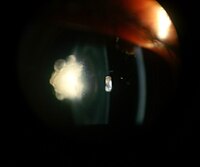
Signs and symptoms


_PHIL_4284_lores.jpg)
Signs and symptoms vary depending on the type of cataract, though considerable overlap occurs. People with
The severity of cataract formation, assuming no other eye disease is present, is judged primarily by a visual acuity test. Other symptoms include frequent changes of glasses and colored halos due to hydration of lens.[citation needed]
Congenital cataracts can result in amblyopia if not treated in a timely manner.[15]
Causes
Age
Age is the most common cause of cataracts.
Oxidative stress associated with lipid peroxidation is an important pathogenic mechanism in cataract formation.[17][18] Senile cataracts are associated with a decrease in antioxidant capacity in the lens.[17] An increase in oxidative stress in the lens or a decrease in the ability to remove reactive oxygen species can lead to the lens becoming more opaque.[17]
Trauma

Blunt trauma causes swelling, thickening, and whitening of the lens fibers. While the swelling normally resolves with time, the white color may remain. In severe blunt trauma, or in injuries that penetrate the eye, the capsule in which the lens sits can be damaged. This damage allows fluid from other parts of the eye to rapidly enter the lens leading to swelling and then whitening, obstructing light from reaching the retina at the back of the eye. Cataracts may develop in 0.7 to 8.0% of cases following electrical injuries.[19] Blunt trauma can also result in star- (stellate) or petal-shaped cataracts.[20]
Radiation
Cataracts can arise as an effect of exposure to various types of radiation. X-rays, one form of
Genetics

The genetic component is strong in the development of cataracts,
Skin diseases
The skin and the lens have the same embryological origin and so can be affected by similar diseases.
Smoking and alcohol
Cigarette smoking has been shown to increase the risk of age-related cataract and nuclear cataract.[25][26] Evidence is conflicting over the effect of alcohol. Some surveys have shown a link, but others which followed people over longer terms have not.[27]
Inadequate vitamin C
Low vitamin C intake and serum levels have been associated with greater cataract rates.[28] However, use of supplements of vitamin C has not demonstrated benefit.[29]
Medications
Some medications, such as systemic, topical, or inhaled
Post-operative
Nearly every person who undergoes a
Hyperbaric oxygen therapy
Other diseases
|
|

|
Diagnosis
Classification

Cataracts may be partial or complete, stationary or progressive, hard or soft. Histologically, the main types of age-related cataracts are nuclear sclerosis, cortical, and posterior subcapsular.[41]
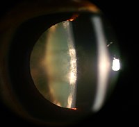
Nuclear sclerosis is the most common type of cataract, and involves the central or 'nuclear' part of the lens. This eventually becomes hard, or 'sclerotic', due to condensation on the lens nucleus and the deposition of brown pigment within the lens. In its advanced stages, it is called a brunescent cataract. In early stages, an increase in sclerosis may cause an increase in refractive index of the lens.[42] This causes a myopic shift (lenticular shift) that decreases hyperopia and enables presbyopic patients to see at near without reading glasses. This is only temporary and is called second sight.[43]
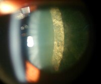
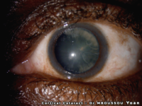
Cortical cataracts are due to the lens cortex (outer layer) becoming opaque. They occur when changes in the fluid contained in the periphery of the lens causes fissuring. When these cataracts are viewed through an
Posterior subcapsular cataracts are cloudy at the back of the lens adjacent to the capsule (or bag) in which the lens sits. Because light becomes more focused toward the back of the lens, they can cause disproportionate symptoms for their size.[43]
An immature cataract has some transparent protein, but with a mature cataract, all the lens protein is opaque. In a hypermature or Morgagnian cataract, the lens proteins have become liquid. Congenital cataract, which may be detected in adulthood, has a different classification and includes lamellar, polar, and sutural cataracts.[44][45]
Cataracts can be classified by using the lens opacities classification system LOCS III. In this system, cataracts are classified based on type as nuclear, cortical, or posterior. The cataracts are further classified based on severity on a scale from 1 to 5. The LOCS III system is highly reproducible.[46]
-
 Posterior polar cataract of an 8-year-old boy in left eye
 -
 Nuclear sclerosis cataract of a 70-year-old male
 -
 Cortical cataract of a 60-year-old male
 -
 Retroillumination of cortical cataract
 -
 Posterior subcapsular cataract of a 16-year-old girl with type 1 diabetes
 -
 Intumescent cataract of a 55-year-old male
 -
 Anterior subcapsular cataract having back shadow
 -
 Posterior subcapsular cataract by retroillumination
 -
 Nuclear sclerosis and posterior polar cataract of a 60-year-old female
 -
 Dense white mature cataract of a 60-year-old male
 -
 Cortical cataract of a melanoderm male


Prevention
Risk factors such as UVB exposure and smoking can be addressed. Although no means of preventing cataracts has been scientifically proven, wearing sunglasses that block ultraviolet light may slow their development.[47][48] While adequate intake of vitamins A, C, and E may protect against the risk of cataracts, clinical trials have shown no benefit from supplements,[29] although the evidence is mixed, but weakly positive, for a potential protective effect of the carotenoids, lutein and zeaxanthin.[49][50][51]
Treatment
Surgical


The appropriateness of surgery depends on a person's particular functional and visual needs and other risk factors.[52] Cataract removal can be performed at any stage and no longer requires ripening of the lens.[clarification needed] Surgery is usually "outpatient" and usually performed using local anesthesia. About 9 of 10 patients can achieve a corrected vision of 20/40 or better after surgery.[42]
Several recent evaluations found that cataract surgery can meet expectations only when significant functional impairment due to cataracts exists before surgery. Visual function estimates such as VF-14 have been found to give more realistic estimates than visual acuity testing alone.[42][53] In some developed countries, a trend to overuse cataract surgery has been noted, which may lead to disappointing results.[54]
Phacoemulsification is the most widely used cataract surgery in the developed world.[55][56] This procedure uses ultrasonic energy to emulsify the cataract lens. Phacoemulsification typically comprises six steps:[citation needed]
- Anaesthetic – The eye is numbed with either a subtenon injection around the eye (see: retrobulbar block) or topical anesthetic eye drops. The former also provides paralysis of the eye muscles.
- Corneal incision – Two cuts are made at the margin of the clear cornea to allow insertion of instruments into the eye.
- Capsulorhexis – A needle or small pair of forceps is used to create a circular hole in the capsule in which the lens sits.
- Phacoemulsification – A handheld ultrasonic probe is used to break up and emulsify the lens into liquid using the energy of ultrasound waves. The resulting 'emulsion' is sucked away.
- Irrigation and aspiration – The cortex, which is the soft outer layer of the cataract, is aspirated or sucked away. Fluid removed is continually replaced with a saline solution to prevent collapse of the structure of the anterior chamber (the front part of the eye).
- Lens insertion – A plastic, foldable lens is inserted into the capsular bag that formerly contained the natural lens. Some surgeons also inject an antibiotic into the eye to reduce the risk of infection. The final step is to inject salt water into the corneal wounds to cause the area to swell and seal the incision.
A
Intracapsular cataract extraction (ICCE) is rarely performed.[59] The lens and surrounding capsule are removed in one piece through a large incision while pressure is applied to the vitreous membrane.[clarification needed] The surgery has a high rate of complications.[clarification needed][citation needed]
Prognosis
Postoperative care

.jpg)
The postoperative recovery period (after removing the cataract) is usually short. The patient is usually ambulatory on the day of surgery, but is advised to move cautiously and avoid straining or heavy lifting for about a month. The eye is usually patched on the day of surgery and use of an eye shield at night is often suggested for several days after surgery.[52]
In all types of surgery, the cataractous lens is removed and replaced with an artificial lens, known as an intraocular lens, which stays in the eye permanently. Intraocular lenses are usually monofocal, correcting for either distance or near vision. Multifocal lenses may be implanted to improve near and distance vision simultaneously, but these lenses may increase the chance of unsatisfactory vision.[16]
Complications
Serious complications of cataract surgery include retinal detachment and endophthalmitis.[60] In both cases, patients notice a sudden decrease in vision. In endophthalmitis, patients often describe pain. Retinal detachment frequently presents with unilateral visual field defects, blurring of vision, flashes of light, or floating spots.[citation needed]
The risk of retinal detachment was estimated as about 0.4% within 5.5 years, corresponding to a 2.3-fold risk increase compared to naturally expected incidence, with older studies reporting a substantially higher risk. The incidence is increasing over time in a somewhat linear manner, and the risk increase lasts for at least 20 years after the procedure. Particular risk factors are younger age, male sex, longer axial length, and complications during surgery. In the highest risk group of patients, the incidence of pseudophakic retinal detachment may be as high as 20%.[61]
The risk of endophthalmitis occurring after surgery is less than one in 1000.[62]
Corneal edema and cystoid macular edema are less serious but more common, and occur because of persistent swelling at the front of the eye in corneal edema or back of the eye in cystoid macular edema.[63] They are normally the result of excessive inflammation following surgery, and in both cases, patients may notice blurred, foggy vision. They normally improve with time and with application of anti-inflammatory drops. The risk of either occurring is around one in 100. It is unclear whether NSAIDs or corticosteroids are superior at reducing postoperative inflammation.[64]
Posterior capsular opacification, also known as after-cataract, is a condition in which months or years after successful cataract surgery, vision deteriorates or problems with glare and light scattering recur, usually due to thickening of the back or posterior capsule surrounding the implanted lens, so-called 'posterior lens capsule opacification'. Growth of natural lens cells remaining after the natural lens was removed may be the cause, and the younger the patient, the greater the chance of this occurring. Management involves cutting a small, circular area in the posterior capsule with targeted beams of energy from a laser, called Nd:YAG laser capsulotomy, after the type of laser used. The laser can be aimed very accurately, and the small part of the capsule which is cut falls harmlessly to the bottom of the inside of the eye. This procedure leaves sufficient capsule to hold the lens in place, but removes enough to allow light to pass directly through to the retina. Serious side effects are rare.[65] Posterior capsular opacification is common and occurs following up to one in four operations, but these rates are decreasing following the introduction of modern intraocular lenses together with a better understanding of the causes.[citation needed]
Vitreous touch syndrome is a possible complication of intracapsular cataract extraction.[66]
Epidemiology
| no data <90 90–180 180–270 270–360 360–450 450–540 | 540–630 630–720 720–810 810–900 900–990 >990 |
Age-related cataracts are responsible for 51% of world blindness, about 20 million people.[68] Globally, cataracts cause moderate to severe disability in 53.8 million (2004), 52.2 million of whom are in low and middle income countries.[69]
In many countries, surgical services are inadequate, and cataracts remain the leading cause of blindness.[68] Even where surgical services are available, low vision associated with cataracts may still be prevalent as a result of long waits for, and barriers to, surgery, such as cost, lack of information and transportation problems.
In the United States, age-related lens changes have been reported in 42% between the ages of 52 and 64,[70] 60% between the ages 65 and 74,[71] and 91% between the ages of 75 and 85.[70] Cataracts affect nearly 22 million Americans age 40 and older. By age 80, more than half of all Americans have cataracts. Direct medical costs for cataract treatment are estimated at $6.8 billion annually.[72]
In the eastern Mediterranean region, cataracts are responsible for over 51% of blindness. Access to eye care in many countries in this region is limited.[73] Childhood-related cataracts are responsible for 5–20% of world childhood blindness.[74]
Vision loss due to cataracts increases the risk of dementia in the elderly population, increases the likelihood of falls and road traffic accidents, and by detrimental effects on the quality of life increases mortality.[75]
History
Cataract surgery was first described by the
In 1468
Etymology
"Cataract" is derived from the Latin cataracta, meaning "waterfall", and from the Ancient Greek καταρράκτης (katarrhaktēs), "down-rushing",[81] from καταράσσω (katarassō) meaning "to dash down"[82] (from kata-, "down"; arassein, "to strike, dash").[83][84] As rapidly running water turns white, so the term may have been used metaphorically to describe the similar appearance of mature ocular opacities. In Latin, cataracta had the alternative meaning "portcullis"[85] and the name possibly passed through French to form the English meaning "eye disease" (early 15th century), on the notion of "obstruction".[86] Early Persian physicians called the term nazul-i-ah, or "descent of the water"—vulgarised into waterfall disease or cataract—believing such blindness to be caused by an outpouring of corrupt humour into the eye.[87]
Research
See also
- Galactosemic cataract – medical condition
- Intraocular lens – Lens implanted in the eye to treat cataracts or myopia
References
- ^ a b c d e f g h i j k l m n o p q r s t "Facts About Cataract". September 2009. Archived from the original on 24 May 2015. Retrieved 24 May 2015.
- ^ S2CID 205670956.
- ^ a b "Visual impairment and blindness Fact Sheet N°282". August 2014. Archived from the original on 12 May 2015. Retrieved 23 May 2015.
- ^ a b c d e "Priority eye diseases". Archived from the original on 24 May 2015. Retrieved 24 May 2015.
- S2CID 195680440.
- PMID 27733282.
- ^ ISBN 978-0-7817-4307-5. Archivedfrom the original on 2015-05-24.
- PMID 16840470.
- ^ Global Data on Visual Impairments 2010 (PDF). WHO. 2012. p. 6. Archived (PDF) from the original on 2015-03-31.
- ^ "Recognizing Cataracts". NIH News in Health. 2017-05-30. Retrieved 2020-02-02.
Try wearing sunglasses or a hat with a brim. Researchers also believe that good nutrition can help reduce the risk of age-related cataract. They recommend eating plenty of green leafy vegetables, fruits, nuts and other healthy foods.
- S2CID 22760161.
- ^ S2CID 205670997.
- ^ a b "Cataract Data and Statistics". National Eye Institute. Retrieved 2019-11-18.
- ^ "Posterior Supcapsular Cataract". Digital Reference of Ophthalmology. Edward S. Harkness Eye Institute, Department of Ophthalmology of Columbia University. 2003. Archived from the original on 27 March 2013. Retrieved 2 April 2013.
- PMID 31317088.
- ^ ISBN 978-0-323-04332-8.[page needed]
- ^ PMID 35163178.
- PMID 24379787.
- S2CID 45814684.
- PMID 27144871.
- ^ PMID 3068822.
- S2CID 8242055.
- ISBN 978-3-8055-7578-2
- ISBN 978-0-323-04332-8
- PMID 22599585.)
{{cite journal}}: CS1 maint: multiple names: authors list (link - S2CID 155984835.)
{{cite journal}}: CS1 maint: multiple names: authors list (link - PMID 18929762.
- S2CID 42785248.
- ^ PMID 22696344.
- S2CID 43843511.
- ^ PMID 8654515.
- PMID 18458771.
- PMID 9927876.
- ^ "Triperanol". MeSH. National Library of Medicine. Archived from the original on 2015-12-22. Retrieved 2013-02-06.
- S2CID 31308270.
- PMID 26891415.
- PMID 24877085.
- PMID 19365028.
- PMID 24406418.
- PMID 29261974. Retrieved 27 February 2023.
- PMID 29356473, retrieved 2023-04-24
- ^ S2CID 27022598.
- ^ ISBN 978-0-12-374203-2, retrieved 2024-02-20
- PMID 14303339.
- PMID 758890.
- ISBN 978-0-323-05751-6.
- S2CID 40041207.
- PMID 8724222. Cited in Five-Year Agenda for the National Eye Health Education Program (NEHEP), p. B-2; National Eye Institute, U.S. National Institutes of Health
- S2CID 206965363.
- S2CID 13634941.
- ISBN 978-1-4939-1934-5.
- ^ ISBN 978-0-07-163420-5.[page needed]
- PMID 22943071.

- S2CID 37414146.
- ISBN 978-1-4939-1935-2
- ]
- PMID 28931202.
- ISBN 978-1-55642-802-9
- ISBN 978-93-5090-274-5
- ISBN 978-3-540-68366-7
- S2CID 260192934.
- PMID 21782099.
- ISBN 978-0-323-37802-4
- PMID 28670710.
- ^ "Posterior capsule opacification – why laser treatment is sometimes needed following cataract surgery". rnib.org.uk. 2014-02-19. Archived from the original on 2009-09-17.
- ^ Banerjee K (2006). "A review and clinical evaluation of per-operative and post-operative complications in case of manual small incision cataract surgery and extracapsular cataract extraction with posterior chamber intra-ocular lens implantation" (PDF). Archived from the original (PDF) on 5 June 2014. Retrieved 1 June 2014.
- ^ "Death and DALY estimates for 2004 by cause for WHO Member States" (xls). World Health Organization. who.int. 2004.
- ^ a b "Priority eye diseases: Cataract". Prevention of Blindness and Visual Impairment. World Health Organization. Archived from the original on 2015-05-24.
- ISBN 978-92-4-156371-0.
- ^ PMID 7395962.
- PMID 879158.
- ^ "Eye Health Statistics at a Glance" (PDF). Archived from the original (PDF) on March 17, 2015.
- ^ "Health Topics: Cataract". World Health Organization – Eastern Mediterranean Regional Office. Archived from the original on 2013-09-27.
- S2CID 208790600.
- PMID 36369026.
- ISBN 978-93-5090-274-5.
- OL 5225311W.
- ^ Elliott J (February 9, 2008). "The Romans carried out cataract ops". BBC News. Archived from the original on February 18, 2008.
- PMC 1034789.
- ISBN 978-0-19-514694-3.
- ^ Liddell HG, Scott R. "καταρράκτης". A Greek-English Lexicon. Archived from the original on 2012-04-05 – via Perseus.
- ^ Liddell HG, Scott R. "καταράσσω". A Greek-English Lexicon. Archived from the original on 2012-04-04 – via Perseus.
- ^ "cataract". Dictionary.com. Dictionary.com, LLC. Retrieved 1 April 2020.
- ^ "cataract". Oxford Dictionaries. Oxford University Press. Archived from the original on 8 October 2012. Retrieved 1 April 2020.
- ^ Lewis CT, Short C. "cataracta". A Latin Dictionary. Archived from the original on 2012-04-04 – via Perseus.
- ^ "cataract". Online Etymology Dictionary. Archived from the original on 2007-10-14.
- ^ Mistaken Science – Topic Powered by eve community Archived 2008-06-22 at the Wayback Machine, Wordcraft Forums, wordcraft.infopop.cc
- PMID 16939459.
- PMID 17162883.
- S2CID 43125880.
- The authors declare a financial interest in a company producing femtosecond laser equipment.
- ^ a b "Stem cells used to repair children's eyes after cataracts". NHS. March 10, 2016. Archived from the original on 11 March 2016. Retrieved 11 March 2016.
Further reading
- Truscott RJ, Friedrich MG (December 2019). "Molecular Processes Implicated in Human Age-Related Nuclear Cataract". Investigative Ophthalmology & Visual Science. 60 (15): 5007–5021. PMID 31791064.
External links
- Cataract at Curlie
- Pictures of different types of cataracts Archived 2013-03-27 at the Wayback Machine