
Cerebral peduncle
| Cerebral peduncle | |
|---|---|
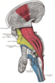
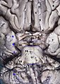
 Superficial dissection of brain-stem. Ventral view. ("cerebral peduncle" visible in red at center-right) | |
 Obtuse section (perpendicular to the brainstem) through superior colliculus showing path of oculomotor nerve (crus cerebri labeled at lower left). | |
| Details | |
| Identifiers | |
| Latin | pedunculus cerebri |
| MeSH | D065850 |
| NeuroNames | 487 |
| NeuroLex ID | birnlex_1202 |
| TA98 | A14.1.06.004 |
| TA2 | 5878 |
| FMA | 62394 |
| Anatomical terms of neuroanatomy | |
The cerebral peduncles are the two stalks that attach the
The cerebral peduncles are located on either side of the midbrain and are the frontmost part of the midbrain, and act as the connectors between the rest of the
ImportantStructure
The descending upper fibers from the internal capsule continue on through the midbrain and are then seen as the fibers in the cerebral peduncles.[5] The corticopontine fibers are found in the outer and inner third of the cerebral peduncle, these are the cortical input to the pontine nuclei.[6] The corticobulbar and corticospinal fibers are found in the middle third of the cerebral peduncle.[7] The corticospinal tract exits the internal capsule and is seen in the mid portion of the cerebral peduncles.
Cranial nerves
Cranial nerve 3 (oculomotor nerve) appears ventrally between the two cerebral peduncles in the interpeduncular fossa. Cranial nerve 4 (trochlear nerve) wraps around the lowest part of the cerebral peduncle.[8]
Additional images
-
 Scheme showing the connections of the several parts of the brain
 -
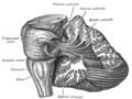
 Deep dissection of brain-stem (lateral view)
 -
 Dissection showing the projection fibers of the cerebellum
 -
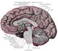 Median sagittal section of brain
 -
 The left optic nerve and the optic tracts
 -
 Upper part of medulla spinalis and hind- and mid-brains; posterior aspect, exposed in situ
 -
 Human brainstem anterior view


See also
- List of regions in the human brain
- Efferent nerve fiber
- Motor neuron (efferent neuron)
- Motor nerve
References
- ISBN 9780071222075.
- ^ Saladin, Kenneth (2010), Anatomy & Physiology The Unity of Form and Function, New York, NY: McGraw-Hill Companies, Inc.
- ^ Swenson, Rand. Review of Clinical and Functional Neuroscience (online ed.). Chapter 8B - Cerebellar Systems: Swenson 2006.
{{cite book}}: CS1 maint: location (link) - ^ HENDELMAN, WALTER J. Atlas of Functional Neuroanatomy (PDF). CRC Press LLC. Archived from the original (PDF) on 8 December 2015. Retrieved 26 November 2015.
- ^ HENDELMAN, WALTER J. Atlas of Functional Neuroanatomy (PDF). CRC Press LLC. Archived from the original (PDF) on 8 December 2015. Retrieved 26 November 2015.
- ^ HENDELMAN, WALTER J. Atlas of Functional Neuroanatomy (PDF). CRC Press LLC. Archived from the original (PDF) on 8 December 2015. Retrieved 26 November 2015.
- ^ HENDELMAN, WALTER J. Atlas of Functional Neuroanatomy (PDF). CRC Press LLC. Archived from the original (PDF) on 8 December 2015. Retrieved 26 November 2015.
- ^ HENDELMAN, WALTER J. Atlas of Functional Neuroanatomy (PDF). CRC Press LLC. Archived from the original (PDF) on 8 December 2015. Retrieved 26 November 2015.