
Thrombus
| Thrombus | |
|---|---|
| Other names | Blood clot |
tinzaparin, unfractionated heparin | |
| Medication | apixaban, edoxaban, and rivaroxaban |
| Deaths | 100,000–300,000 each year |
A thrombus (pl.: thrombi), colloquially called a blood clot, is the final product of the
In the
Mural thrombi are thrombi that adhere to the wall of a large
Classification
Thrombi are classified into two major groups depending on their location and the relative amount of platelets and red blood cells (RBCs).[4] The two major groups are:
- Arterial or white thrombi (characterized by predominance of platelets)
- Venous or red thrombi (characterized by predominance of red blood cells).
Microclots
In the
Microclots can cause a number of problems particularly affecting the alveoli in the lungs of the respiratory system, resulting from reduced oxygen supply. Microclots have been found to be a characteristic feature in severe cases of COVID-19, and in long COVID.[1][6]
Mural thrombi
Mural thrombi are thrombi that form and adhere on the inner wall of a large
A mural thrombus can affect any heart chamber. When found in the
Cause

It was suggested over 150 years ago that thrombus formation is a result of abnormalities in blood flow, vessel wall, and blood components. This concept is now known as Virchow's triad. The three factors have been further refined to include circulatory stasis, vascular wall injury, and hypercoagulable state, all of which contribute to increased risk for venous thromboembolism and other cardiovascular diseases.[4]
Virchow's triad describes the pathogenesis of thrombus formation:[7][8]
- Endothelial injury: Injury to the endothelium (interior surface of blood vessel), causing platelet activation and aggregation;
- Common causes include: trauma, smoking, hypertension, atheroma.
- Hemodynamic changes (stasis, turbulence): Blood stasis promotes greater contact between platelets/coagulative factors with vascular endothelium. If rapid blood circulation (e.g., because of tachycardia) occurs within vessels that have endothelial injuries, that creates disordered flow (turbulence) that can lead to the formation of thrombosis;[9]
- Common causes of stasis include anything that leads to prolonged immobility and reduced blood flow such as: trauma/broken bones and extended air travel.
- Hypercoagulability (also called thrombophilia; any disorder of the blood that predisposes to thrombosis);[10]
- Common causes include: cancer () – prevents Factor V inactivation leading to increased coagulability.
Pathophysiology

A thrombus occurs when the hemostatic process, which normally occurs in response to injury, becomes activated in an uninjured or slightly injured vessel. A thrombus in a large blood vessel will decrease blood flow through that vessel (termed a mural thrombus). In a small blood vessel, blood flow may be completely cut off (termed an occlusive thrombus), resulting in death of tissue supplied by that vessel. If a thrombus dislodges and becomes free-floating, it is considered an embolus.[citation needed] If an embolus becomes trapped within a blood vessel, it blocks blood flow and is termed as an embolism. Embolisms, depending on their specific location, can cause more significant effects like strokes, heart attacks, or even death.[11]

Some of the conditions which increase the risk of blood clots developing include
Formation
Platelet activation occurs through injuries that damage the
-

-
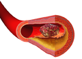 Illustration depicting thrombus formation over arterial plaque.
 -
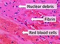 Composition of a fresh thrombus at microscopy, showing nuclear debris in a background of fibrin and red blood cells.


Prevention
Anticoagulants are drugs used to prevent the formation of blood clots, reducing the risk of stroke, heart attack and pulmonary embolism. Heparin and warfarin are used to inhibit the formation and growth of existing thrombi, with the former used for acute anticoagulation while the latter is used for long-term anticoagulation.[8] The mechanism of action of heparin and warfarin are different as they work on different pathways of the coagulation cascade.[13]
Heparin works by binding to and activating the enzyme inhibitor antithrombin III, an enzyme that acts by inactivating thrombin and factor Xa.[13] In contrast, warfarin works by inhibiting vitamin K epoxide reductase, an enzyme needed to synthesize vitamin K dependent clotting factors II, VII, IX, and X.[13][14] Bleeding time with heparin and warfarin therapy can be measured with the partial thromboplastin time (PTT) and prothrombin time (PT), respectively.[14]
Treatment
Once clots have formed, other drugs can be used to promote
There are also some anticoagulants that come from animals that work by dissolving fibrin. For example, Haementeria ghilianii, an Amazon leech, produces an enzyme called hementin from its salivary glands.[18]
Prognosis
Thrombus formation can have one of four outcomes: propagation, embolization, dissolution, and organization and recanalization.[19]
- Propagation of a thrombus occurs towards the direction of the heart and involves the accumulation of additional platelets and fibrin. This means that it is anterograde in veins or retrograde in arteries.
- Embolization occurs when the thrombus breaks free from the vascular wall and becomes mobile, thereby traveling to other sites in the vasculature. A venous embolus (mostly from deep vein thrombosis in the lower limbs) will travel through the systemic circulation, reach the right side of the heart, and travel through the pulmonary artery, resulting in a pulmonary embolism. Arterial thrombosis resulting from hypertension or atherosclerosis can become mobile and the resulting emboli can occlude any artery or arteriole downstream of the thrombus formation. This means that cerebral stroke, myocardial infarction, or any other organ can be affected.
- Dissolution occurs when the fibrinolytic mechanisms break up the thrombus and blood flow is restored to the vessel. This may be aided by fibrinolytic drugs such as Tissue Plasminogen Activator (tPA) in instances of coronary artery occlusion. The best response to fibrinolytic drugs is within a couple of hours, before the fibrin meshwork of the thrombus has been fully developed.
- Organization and recanalization involves the ingrowth of smooth muscle cells, fibroblasts and endothelium into the fibrin-rich thrombus. If recanalization proceeds it provides capillary-sized channels through the thrombus for continuity of blood flow through the entire thrombus but may not restore sufficient blood flow for the metabolic needs of the downstream tissue.[7]
See also
- Thrombogenicity (the tendency to clot)
- National Blood Clot Alliance
- Hemorrhoid
References
- ^ PMID 34425843.
- ^ PMID 30484999. Retrieved 11 February 2022.
- ^ PMID 28856146.
- ^ a b "Thrombus Formation – Virchow's triad & Types of Thrombi". Thrombosis Adviser. Bayer AG. Retrieved 20 March 2020.
- ^ "Medical Definition of micro thrombus". www.merriam-webster.com. Retrieved 22 February 2023.
- S2CID 255608747.
- ^ OCLC 879416939.
- ^ a b "Venous thromboembolism (VTE) | McMaster Pathophysiology Review". www.pathophys.org. 26 September 2012. Retrieved 2018-11-03.
- PMID 30969519, retrieved 2020-06-18
- PMID 19880774.
- ^ Marieb, Elaina N. Human Anatomy and Physiology (11th ed.). Pearson.
- PMID 18753650.
- ^ PMID 25671002.
- ^ OCLC 881019575.
- ISBN 978-0-07-337825-1.
- PMID 27677179.
- PMID 21707475.
- PMID 1772982.
- ISBN 978-1-4160-2973-1.
External links
- Muscle Relaxing Drugs Can Reduce Lethal Blood Clots Archived 2009-02-04 at the Wayback Machine
- Air Pollution Triggers Blood Clots – US Study.
| Authority control databases: National |
|---|