NADPH while freeing oxygen from water in the cells. The ATP and NADPH is then used to make organic molecules from carbon dioxide in a process known as the Calvin cycle. Chloroplasts carry out a number of other functions, including fatty acid synthesis, amino acid synthesis, and the immune response in plants. The number of chloroplasts per cell varies from one, in unicellular algae, up to 100 in plants like Arabidopsis and wheat
Chloroplasts cannot be made by the plant cell and must be inherited by each daughter cell during cell division.
With one exception (the
tertiary endosymbiotic events
.
The word chloroplast is derived from the Greek words chloros (χλωρός), which means green, and plastes (πλάστης), which means "the one who forms".[4]
Discovery
The first definitive description of a chloroplast (Chlorophyllkörnen, "grain of chlorophyll") was given by Hugo von Mohl in 1837 as discrete bodies within the green plant cell.[5] In 1883, Andreas Franz Wilhelm Schimper named these bodies as "chloroplastids" (Chloroplastiden).[6] In 1884, Eduard Strasburger adopted the term "chloroplasts" (Chloroplasten).[7][8][9]
Lineages and evolution
Chloroplasts are one of many types of organelles in the plant cell. They are considered to have evolved from
Chloroplasts are considered endosymbiotic Cyanobacteria.[14] Cyanobacteria are sometimes called blue-green algae even though they are prokaryotes. They are a diverse phylum of gram-negativebacteria capable of carrying out photosynthesis. Cyanobacteria also contain a peptidoglycan cell wall, which is thicker than in other gram-negative bacteria, and which is located between their two cell membranes.[15] Like chloroplasts, they have thylakoids within them.[16] On the thylakoid membranes are photosynthetic pigments, including chlorophyll a.[17]Phycobilins are also common cyanobacterial pigments, usually organized into hemispherical phycobilisomes attached to the outside of the thylakoid membranes (phycobilins are not shared with all chloroplasts though).[17][18]
endosymbiosis, or "cell living inside another cell with a mutual benefit for both". The external cell is commonly referred to as the host while the internal cell is called the endosymbiont.[10]
Chloroplasts are believed to have arisen after
serial endosymbiosis—an early eukaryote engulfing the mitochondrion ancestor, and some descendants of it then engulfing the chloroplast ancestor, creating a cell with both chloroplasts and mitochondria.[10]
Whether or not primary chloroplasts came from a single endosymbiotic event, or many independent engulfments across various eukaryotic lineages, has long been debated. It is now generally held that organisms with primary chloroplasts share
cyanobacterium 90–500 million years ago.[32][31][22]
These chloroplasts, which can be traced back directly to a cyanobacterial ancestor, are known as primary
Usually the endosymbiosis event is considered to have occurred in the
carbon fixation enzymeRuBisCO in. The starch that they synthesize collects outside the chloroplast.[17] Like cyanobacteria, glaucophyte and rhodophyte chloroplast thylakoids are studded with light collecting structures called phycobilisomes.[17][33] For these reasons, glaucophyte chloroplasts are considered a primitive intermediate between cyanobacteria and the more evolved chloroplasts in red algae and plants.[33]
rhodophyte, or red algae chloroplast group is another large and diverse chloroplast lineage.[19] Rhodophyte chloroplasts are also called rhodoplasts,[33] literally "red chloroplasts".[37]
Rhodoplasts have a double membrane with an intermembrane space and phycobilin pigments organized into phycobilisomes on the thylakoid membranes, preventing their thylakoids from stacking.[17] Some contain pyrenoids.[33] Rhodoplasts have chlorophyll a and phycobilins[35] for photosynthetic pigments; the phycobilin phycoerythrin is responsible for giving many red algae their distinctive red color.[36] However, since they also contain the blue-green chlorophyll a and other pigments, many are reddish to purple from the combination.[33][dubious – discuss] The red phycoerytherin pigment is an adaptation to help red algae catch more sunlight in deep water[33]—as such, some red algae that live in shallow water have less phycoerythrin in their rhodoplasts, and can appear more greenish.[36] Rhodoplasts synthesize a form of starch called floridean starch,[33] which collects into granules outside the rhodoplast, in the cytoplasm of the red alga.[17]
Hæmatococcus pluvialis, due to accessory pigments that override the chlorophylls' green colors. Chloroplastida chloroplasts have lost the peptidoglycan wall between their double membrane, leaving an intermembrane space.[17] Some plants seem to have kept the genes for the synthesis of the peptidoglycan layer, though they've been repurposed for use in chloroplast division instead.[39]
Most of the chloroplasts depicted in this article are green chloroplasts.
Green algae and plants keep their starchinside their chloroplasts,[17][35][38] and in plants and some algae, the chloroplast thylakoids are arranged in grana stacks. Some green algal chloroplasts contain a structure called a pyrenoid,[17] which is functionally similar to the glaucophyte carboxysome in that it is where RuBisCO and CO2 are concentrated in the chloroplast.[40]
, a green alga that contains a pyrenoid surrounded by starch.
Helicosporidium is a genus of nonphotosynthetic parasitic green algae that is thought to contain a vestigial chloroplast.[35] Genes from a chloroplast[41] and nuclear genes indicating the presence of a chloroplast have been found in Helicosporidium[35] even if nobody's seen the chloroplast itself.[35]
Paulinella chromatophora
While most chloroplasts originate from that first set of endosymbiotic events, Paulinella chromatophora is an exception that acquired a photosynthetic cyanobacterial endosymbiont more recently. It is not clear whether that symbiont is closely related to the ancestral chloroplast of other eukaryotes.[19] Being in the early stages of endosymbiosis, Paulinella chromatophora can offer some insights into how chloroplasts evolved.[26][42]Paulinella cells contain one or two sausage-shaped blue-green photosynthesizing structures called chromatophores,[26][42] descended from the cyanobacterium Synechococcus. Chromatophores cannot survive outside their host.[26] Chromatophore DNA is about a million base pairs long, containing around 850 protein-encoding genes—far less than the three million base pair Synechococcus genome,[26] but much larger than the approximately 150,000 base pair genome of the more assimilated chloroplast.[43][44][45] Chromatophores have transferred much less of their DNA to the nucleus of their host. About 0.3–0.8% of the nuclear DNA in Paulinella is from the chromatophore, compared with 11–14% from the chloroplast in plants.[42]
Many other organisms obtained chloroplasts from the primary chloroplast lineages through secondary endosymbiosis—engulfing a red or green alga that contained a chloroplast. These chloroplasts are known as secondary plastids.[33]
While primary chloroplasts have a double membrane from their
phagosomal vacuole from the host's cell membrane.[19]
Secondary endosymbiosis consisted of a eukaryoticalga being engulfed by another eukaryote, forming a chloroplast with three or four membranes.
Diagram of a four membraned chloroplast containing a nucleomorph.
The genes in the phagocytosed eukaryote's nucleus are often transferred to the secondary host's nucleus.[19]Cryptomonads and chlorarachniophytes retain the phagocytosed eukaryote's nucleus, an object called a nucleomorph,[19] located between the second and third membranes of the chloroplast.[17][27]
All secondary chloroplasts come from
glaucophytes have been observed, probably because glaucophytes are relatively rare in nature, making them less likely to have been taken up by another eukaryote.[19]
Green algal derived chloroplasts
alveolates, stramenopiles and haptophytes)[47] in three or four separate engulfments.[48] Many green algal derived chloroplasts contain pyrenoids, but unlike chloroplasts in their green algal ancestors, storage product collects in granules outside the chloroplast.[17]
euglenophyte
, contains secondary chloroplasts from green algae.
Euglenophytes
Euglenophytes are a group of common
Euglenophyte chloroplasts have three membranes—it is thought that the membrane of the primary endosymbiont was lost, leaving the cyanobacterial membranes, and the secondary host's phagosomal membrane.[19] Euglenophyte chloroplasts have a pyrenoid and thylakoids stacked in groups of three. Photosynthetic product is stored in the form of paramylon, which is contained in membrane-bound granules in the cytoplasm of the euglenophyte.[17][35]
green alga
.
Chlorarachniophytes
Chlorarachniophytes /ˌklɔːrəˈræknioʊˌfaɪts/ are a rare group of organisms that also contain chloroplasts derived from green algae,[19] though their story is more complicated than that of the euglenophytes. The ancestor of chlorarachniophytes is thought to have been a eukaryote with a red algal derived chloroplast. It is then thought to have lost its first red algal chloroplast, and later engulfed a green alga, giving it its second, green algal derived chloroplast.[35]
Chlorarachniophyte chloroplasts are bounded by four membranes, except near the cell membrane, where the chloroplast membranes fuse into a double membrane.[17] Their thylakoids are arranged in loose stacks of three.[17] Chlorarachniophytes have a form of polysaccharide called chrysolaminarin, which they store in the cytoplasm,[35] often collected around the chloroplast pyrenoid, which bulges into the cytoplasm.[17]
Chlorarachniophyte chloroplasts are notable because the green alga they are derived from has not been completely broken down—its nucleus still persists as a nucleomorph[19] found between the second and third chloroplast membranes[17]—the periplastid space, which corresponds to the green alga's cytoplasm.[35]
Prasinophyte-derived dinophyte chloroplast
green alga containing a primary chloroplast (making a secondary chloroplast).[35]
Red algal derived chloroplasts
Cryptophytes
rough endoplasmic reticulum. They synthesize ordinary starch, which is stored in granules found in the periplastid space—outside the original double membrane, in the place that corresponds to the red alga's cytoplasm. Inside cryptophyte chloroplasts is a pyrenoid and thylakoids in stacks of two.[17]
Their chloroplasts do not have phycobilisomes,[17] but they do have phycobilin pigments which they keep in their thylakoid space, rather than anchored on the outside of their thylakoid membranes.[17][19]
Cryptophytes may have played a key role in the spreading of red algal based chloroplasts.[50][51]
Haptophytes are similar and closely related to cryptophytes or heterokontophytes.[35] Their chloroplasts lack a nucleomorph,[17][19] their thylakoids are in stacks of three, and they synthesize chrysolaminarin sugar, which they store completely outside of the chloroplast, in the cytoplasm of the haptophyte.[17]
Heterokont chloroplasts are very similar to haptophyte chloroplasts, containing a pyrenoid, triplet thylakoids, and with some exceptions,[17] having four layer plastidic envelope, the outermost epiplastid membrane connected to the endoplasmic reticulum. Like haptophytes, heterokontophytes store sugar in chrysolaminarin granules in the cytoplasm.[17] Heterokontophyte chloroplasts contain chlorophyll a and with a few exceptions[17]chlorophyll c,[19] but also have carotenoids which give them their many colors.[36]
Apicomplexans, chromerids, and dinophytes
The alveolates are a major clade of unicellular eukaryotes of both autotrophic and heterotrophic members. The most notable shared characteristic is the presence of cortical (outer-region) alveoli (sacs). These are flattened vesicles (sacs) packed into a continuous layer just under the membrane and supporting it, typically forming a flexible pellicle (thin skin). In dinoflagellates they often form armor plates. Many members contain a red-algal derived plastid. One notable characteristic of this diverse group is the frequent loss of photosynthesis. However, a majority of these heterotrophs continue to process a non-photosynthetic plastid.[52]
Apicomplexans
vestigial red algal derived chloroplast[53][35] called an apicoplast, which they inherited from their ancestors. Other apicomplexans like Cryptosporidium have lost the chloroplast completely.[53] Apicomplexans store their energy in amylopectin granules that are located in their cytoplasm, even though they are nonphotosynthetic.[17]
Apicoplasts have lost all photosynthetic function, and contain no photosynthetic pigments or true thylakoids. They are bounded by four membranes, but the membranes are not connected to the
iron-sulfur clusters, and carry out part of the heme pathway.[53] This makes the apicoplast an attractive target for drugs to cure apicomplexan-related diseases.[33] The most important apicoplast function is isopentenyl pyrophosphate synthesis—in fact, apicomplexans die when something interferes with this apicoplast function, and when apicomplexans are grown in an isopentenyl pyrophosphate-rich medium, they dump the organelle.[53]
Chromerids
The
Chromera velia, was discovered and first isolated in 2001. The discovery of Chromera velia with similar structure to the apicomplexans, provides an important link in the evolutionary history of the apicomplexans and dinophytes. Their plastids have four membranes, lack chlorophyll c and use the type II form of RuBisCO obtained from a horizontal transfer event.[54]
Most dinophyte chloroplasts are secondary red algal derived chloroplasts. Many other dinophytes have lost the chloroplast (becoming the nonphotosynthetic kind of dinoflagellate), or replaced it though tertiary endosymbiosis[55]—the engulfment of another eukaryotic algae containing a red algal derived chloroplast. Others replaced their original chloroplast with a green algal derived one.[19][35][49]
Most dinophyte chloroplasts contain form II RuBisCO, at least the
beta-carotene, and at least one dinophyte-unique xanthophyll (peridinin, dinoxanthin, or diadinoxanthin), giving many a golden-brown color.[52][49] All dinophytes store starch in their cytoplasm, and most have chloroplasts with thylakoids arranged in stacks of three.[17]
reduced and fragmented into many small circles. Most of the genome has migrated to the nucleus, and only critical photosynthesis-related genes remain in the chloroplast.[49]
The peridinin chloroplast is thought to be the dinophytes' "original" chloroplast,[49] which has been lost, reduced, replaced, or has company in several other dinophyte lineages.[35]
phagosomal vacuole.[57] However, the haptophyte was heavily reduced, stripped of a few membranes and its nucleus, leaving only its chloroplast (with its original double membrane), and possibly one or two additional membranes around it.[35][57]
genome reduction, and might have even been expanded.[35] Diatoms have been engulfed by dinoflagellates at least three times.[35]
The diatom endosymbiont is bounded by a single membrane,[49] inside it are chloroplasts with four membranes. Like the diatom endosymbiont's diatom ancestor, the chloroplasts have triplet thylakoids and pyrenoids.[58]
In some of these
genera, the diatom endosymbiont's chloroplasts aren't the only chloroplasts in the dinophyte. The original three-membraned peridinin chloroplast is still around, converted to an eyespot.[19][35]
kleptoplast—if so, Dinophysis chloroplasts wear out and Dinophysis species must continually engulf cryptophytes to obtain new chloroplasts to replace the old ones.[49]
glaucophytes, red algae, and other algal groups are extremely underrepresented, potentially introducing some bias in views of "typical" chloroplast DNA structure and content.[66]
Chloroplast DNA Interactive gene map of chloroplast DNA from
introns
.
With few exceptions, most chloroplasts have their entire chloroplast genome combined into a single large circular DNA molecule,[66] typically 120,000–170,000 base pairs long.[43][44][45][21] They can have a contour length of around 30–60 micrometers, and have a mass of about 80–130 million daltons.[67]
While usually thought of as a circular molecule, there is some evidence that chloroplast DNA molecules more often take on a
Many chloroplast DNAs contain two inverted repeats, which separate a long single copy section (LSC) from a short single copy section (SSC).[45]
While a given pair of inverted repeats are rarely completely identical, they are always very similar to each other, apparently resulting from concerted evolution.[66]
The inverted repeats vary wildly in length, ranging from 4,000 to 25,000 base pairs long each and containing as few as four or as many as over 150 genes.[66] Inverted repeats in plants tend to be at the upper end of this range, each being 20,000–25,000 base pairs long.[45][69]
The inverted repeat regions are highly
rhodophyceae), suggesting that they predate the chloroplast,[66] though some chloroplast DNAs have since lost[69][70] or flipped the inverted repeats (making them direct repeats).[66] It is possible that the inverted repeats help stabilize the rest of the chloroplast genome, as chloroplast DNAs which have lost some of the inverted repeat segments tend to get rearranged more.[70]
Nucleoids
New chloroplasts may contain up to 100 copies of their DNA,[43] though the number of chloroplast DNA copies decreases to about 15–20 as the chloroplasts age.[71] They are usually packed into nucleoids, which can contain several identical chloroplast DNA rings. Many nucleoids can be found in each chloroplast.[67]
In primitive red algae, the chloroplast DNA nucleoids are clustered in the center of the chloroplast, while in green plants and green algae, the nucleoids are dispersed throughout the stroma.[72]
Though chloroplast DNA is not associated with true histones,[10] in red algae, similar proteins that tightly pack each chloroplast DNA ring into a nucleoid have been found.[72]
Chloroplast DNA replication via multiple D-loop mechanisms. Adapted from Krishnan NM, Rao BJ's paper "A comparative approach to elucidate chloroplast genome replication."
The mechanism for chloroplast DNA (cpDNA) replication has not been conclusively determined, but two main models have been proposed. Scientists have attempted to observe chloroplast replication via
electron microscopy since the 1970s.[75][76] The results of the microscopy experiments led to the idea that chloroplast DNA replicates using a double displacement loop (D-loop). As the D-loop moves through the circular DNA, it adopts a theta intermediary form, also known as a Cairns replication intermediate, and completes replication with a rolling circle mechanism.[75][68]
Transcription starts at specific points of origin. Multiple replication forks open up, allowing replication machinery to transcribe the DNA. As replication continues, the forks grow and eventually converge. The new cpDNA structures separate, creating daughter cpDNA chromosomes.
In addition to the early microscopy experiments, this model is also supported by the amounts of deamination seen in cpDNA.[75] Deamination occurs when an amino group is lost and is a mutation that often results in base changes. When adenine is deaminated, it becomes hypoxanthine. Hypoxanthine can bind to cytosine, and when the XC base pair is replicated, it becomes a GC (thus, an A → G base change).[77]
Over time, base changes in the DNA sequence can arise from deamination mutations. When adenine is deaminated, it becomes hypoxanthine, which can pair with cytosine. During replication, the cytosine will pair with guanine, causing an A --> G base change.
In cpDNA, there are several A → G deamination gradients. DNA becomes susceptible to deamination events when it is single stranded. When replication forks form, the strand not being copied is single stranded, and thus at risk for A → G deamination. Therefore, gradients in deamination indicate that replication forks were most likely present and the direction that they initially opened (the highest gradient is most likely nearest the start site because it was single stranded for the longest amount of time).[75] This mechanism is still the leading theory today; however, a second theory suggests that most cpDNA is actually linear and replicates through homologous recombination. It further contends that only a minority of the genetic material is kept in circular chromosomes while the rest is in branched, linear, or other complex structures.[75][68]
One of competing model for cpDNA replication asserts that most cpDNA is linear and participates in
bacteriophage T4.[68][78] It has been established that some plants have linear cpDNA, such as maize, and that more species still contain complex structures that scientists do not yet understand.[68] When the original experiments on cpDNA were performed, scientists did notice linear structures; however, they attributed these linear forms to broken circles.[68] If the branched and complex structures seen in cpDNA experiments are real and not artifacts of concatenated circular DNA or broken circles, then a D-loop mechanism of replication is insufficient to explain how those structures would replicate.[68] At the same time, homologous recombination does not expand the multiple A --> G gradients seen in plastomes.[75]
Because of the failure to explain the deamination gradient as well as the numerous plant species that have been shown to have circular cpDNA, the predominant theory continues to hold that most cpDNA is circular and most likely replicates via a D loop mechanism.
Gene content and protein synthesis
The chloroplast genome most commonly includes around 100 genes
Among land plants, the contents of the chloroplast genome are fairly similar.[45]
Chloroplast genome reduction and gene transfer
Over time, many parts of the chloroplast genome were transferred to the
reduced compared to that of free-living cyanobacteria. Chloroplasts may contain 60–100 genes whereas cyanobacteria often have more than 1500 genes in their genome.[81] Recently, a plastid without a genome was found, demonstrating chloroplasts can lose their genome during endosymbiotic the gene transfer process.[82]
Endosymbiotic gene transfer is how we know about the
green algal derived chloroplast at some point, which was subsequently replaced by the red chloroplast.[47]
In land plants, some 11–14% of the DNA in their nuclei can be traced back to the chloroplast,[42] up to 18% in Arabidopsis, corresponding to about 4,500 protein-coding genes.[83] There have been a few recent transfers of genes from the chloroplast DNA to the nuclear genome in land plants.[44]
Of the approximately 3000 proteins found in chloroplasts, some 95% of them are encoded by nuclear genes. Many of the chloroplast's protein complexes consist of subunits from both the chloroplast genome and the host's nuclear genome. As a result,
protein synthesis must be coordinated between the chloroplast and the nucleus. The chloroplast is mostly under nuclear control, though chloroplasts can also give out signals regulating gene expression in the nucleus, called retrograde signaling.[84] Recent research indicates that parts of the retrograde signaling network once considered characteristic for land plants emerged already in an algal progenitor,[85][86][87] integrating into co-expressed cohorts of genes in the closest algal relatives of land plants.[88]
Protein synthesis within chloroplasts relies on two RNA polymerases. One is coded by the chloroplast DNA, the other is of nuclear origin. The two RNA polymerases may recognize and bind to different kinds of promoters within the chloroplast genome.[89] The ribosomes in chloroplasts are similar to bacterial ribosomes.[90]
This section needs expansion with: Genome size differences between algae and land plants, chloroplast stuff coded by the nucleus. You can help by adding to it. (January 2013)
Because so many chloroplast genes have been moved to the nucleus, many proteins that would originally have been translated in the chloroplast are now synthesized in the cytoplasm of the plant cell. These proteins must be directed back to the chloroplast, and imported through at least two chloroplast membranes.[91]
Curiously, around half of the protein products of transferred genes aren't even targeted back to the chloroplast. Many became
topologically outside of the cell because to reach the chloroplast from the cytosol, the cell membrane must be crossed, which signifies entrance into the extracellular space. In those cases, chloroplast-targeted proteins do initially travel along the secretory pathway.[35]
Because the cell acquiring a chloroplast
protein targeting system to avoid having chloroplast proteins being sent to the wrong organelle.[91]
carboxyl group
(CO2H) is at the right.
In most, but not all cases, nuclear-encoded chloroplast proteins are
cleavable transit peptide that's added to the N-terminus of the protein precursor. Sometimes the transit sequence is found on the C-terminus of the protein,[93] or within the functional part of the protein.[91]
Transport proteins and membrane translocons
After a chloroplast
phosphate group to many (but not all) of them in their transit sequences.[91]
Phosphorylation helps many proteins bind the polypeptide, keeping it from folding prematurely.[91] This is important because it prevents chloroplast proteins from assuming their active form and carrying out their chloroplast functions in the wrong place—the cytosol.[95][96] At the same time, they have to keep just enough shape so that they can be recognized by the chloroplast.[95] These proteins also help the polypeptide get imported into the chloroplast.[91]
From here, chloroplast proteins bound for the stroma must pass through two protein complexes—the
Transmission electron microscope image of a chloroplast. Grana of thylakoids
and their connecting lamellae are clearly visible.
In land plants, chloroplasts are generally lens-shaped, 3–10 μm in diameter and 1–3 μm thick.[97][21] Corn seedling chloroplasts are ≈20 µm3 in volume.[21] Greater diversity in chloroplast shapes exists among the algae, which often contain a single chloroplast[17] that can be shaped like a net (e.g., Oedogonium),[98] a cup (e.g., Chlamydomonas),[99] a ribbon-like spiral around the edges of the cell (e.g., Spirogyra),[100] or slightly twisted bands at the cell edges (e.g., Sirogonium).[101] Some algae have two chloroplasts in each cell; they are star-shaped in Zygnema,[102] or may follow the shape of half the cell in orderDesmidiales.[103] In some algae, the chloroplast takes up most of the cell, with pockets for the nucleus and other organelles,[17] for example, some species of Chlorella have a cup-shaped chloroplast that occupies much of the cell.[104]
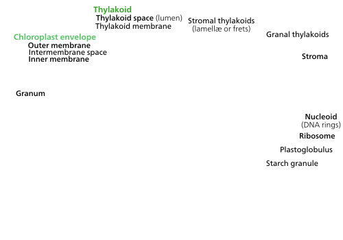
All chloroplasts have at least three membrane systems—the outer chloroplast membrane, the inner chloroplast membrane, and the thylakoid system. Chloroplasts that are the product of secondary endosymbiosis may have additional membranes surrounding these three.[46] Inside the outer and inner chloroplast membranes is the chloroplast stroma, a semi-gel-like fluid[33] that makes up much of a chloroplast's volume, and in which the thylakoid system floats.
Chloroplast ultrastructure(interactive diagram) Chloroplasts have at least three distinct membrane systems, and a variety of things can be found in their stroma.
There are some common misconceptions about the outer and inner chloroplast membranes. The fact that chloroplasts are surrounded by a double membrane is often cited as evidence that they are the descendants of endosymbiotic
cyanobacterium—which is not true—both chloroplast membranes are homologous to the cyanobacterium's original double membranes.[19]
The chloroplast double membrane is also often compared to the
analogous to it is the internal thylakoid system. Even so, in terms of "in-out", the direction of chloroplast H+ ion flow is in the opposite direction compared to oxidative phosphorylation in mitochondria.[33][105] In addition, in terms of function, the inner chloroplast membrane, which regulates metabolite passage and synthesizes some materials, has no counterpart in the mitochondrion.[33]
The outer chloroplast membrane is a semi-porous membrane that small molecules and
TOC complex, or translocon on the outer chloroplast membrane.[91]
The chloroplast membranes sometimes protrude out into the cytoplasm, forming a
dividing chloroplasts.[110] However, there is a growing body of evidence that stromules are functional, integral features of plant cell plastids, not merely artifacts.[111]
Intermembrane space and peptidoglycan wall
peptidoglycan wall
between their inner and outer chloroplast membranes.
Usually, a thin intermembrane space about 10–20
nanometers thick exists between the outer and inner chloroplast membranes.[112]
cyanobacterial ancestors, which is located between their two cell membranes. These chloroplasts are called muroplasts (from Latin "mura", meaning "wall"). Other chloroplasts were assumed to have lost the cyanobacterial wall, leaving an intermembrane space between the two chloroplast envelope membranes,[33] but has since been found also in moss, lycophytes and ferns.[113]
Chloroplast ribosomes Comparison of a chloroplast ribosome (green) and a bacterial ribosome (yellow). Important features common to both ribosomes and chloroplast-unique features are labeled.
Chloroplasts have their own ribosomes, which they use to synthesize a small fraction of their proteins. Chloroplast ribosomes are about two-thirds the size of
bacterial ribosomes,[10] chloroplast translation is more complex than in bacteria, so chloroplast ribosomes include some chloroplast-unique features.[116][117]
Small subunit
Shine-Dalgarno sequence recognition,[118] which is considered essential for translation initiation in most chloroplasts and prokaryotes.[119][120] Such loss is also rarely observed in other plastids and prokaryotes.[118][121] An additional 4.5S rRNA with homology to the 3' tail of 23S is found in "higher" plants.[117]
Plastoglobuli
Plastoglobuli (singularplastoglobulus, sometimes spelled plastoglobule(s)), are spherical bubbles of
etioplasts, but decrease in number as the etioplasts mature into chloroplasts.[122]
Plastoglobuli contain both structural proteins and enzymes involved in
Plastoglobuli were once thought to be free-floating in the stroma, but it is now thought that they are permanently attached either to a thylakoid or to another plastoglobulus attached to a thylakoid, a configuration that allows a plastoglobulus to exchange its contents with the thylakoid network.[122] In normal green chloroplasts, the vast majority of plastoglobuli occur singularly, attached directly to their parent thylakoid. In old or stressed chloroplasts, plastoglobuli tend to occur in linked groups or chains, still always anchored to a thylakoid.[122]
Plastoglobuli form when a bubble appears between the layers of the
amyloplasts, they can be big enough to distort the shape of the organelle.[112] Starch granules are simply accumulations of starch in the stroma, and are not bounded by a membrane.[112]
Starch granules appear and grow throughout the day, as the chloroplast synthesizes
sugars, and are consumed at night to fuel respiration and continue sugar export into the phloem,[124] though in mature chloroplasts, it is rare for a starch granule to be completely consumed or for a new granule to accumulate.[123]
Starch granules vary in composition and location across different chloroplast lineages. In
mesophyll chloroplasts, which do not synthesize sugars, lack starch granules.[33]
hornworts[127] and algae contain structures called pyrenoids. They are not found in higher plants.[128] Pyrenoids are roughly spherical and highly refractive bodies which are a site of starch accumulation in plants that contain them. They consist of a matrix opaque to electrons, surrounded by two hemispherical starch plates. The starch is accumulated as the pyrenoids mature.[129] In algae with carbon concentrating mechanisms, the enzyme RuBisCO is found in the pyrenoids. Starch can also accumulate around the pyrenoids when CO2 is scarce.[128] Pyrenoids can divide to form new pyrenoids, or be produced "de novo".[129][130]
Thylakoid system
Scanning transmission electron microscope imaging of a chloroplast (Top) 10-nm-thick STEM tomographic slice of a lettuce chloroplast. Grana stacks are interconnected by unstacked stromal thylakoids, called "stroma lamellae". Round inclusions associated with the thylakoids are plastoglobules. Scalebar=200 nm. See.[131] (Bottom) Large-scale 3D model generated from segmentation of tomographic reconstructions by STEM. grana=yellow; stroma lamellae=green; plastoglobules=purple; chloroplast envelope=blue. See.[131]
algal chloroplasts, the thylakoids are free floating.[17]
Thylakoid structure
Granum-stroma assembly structure The prevailing model of the granum-stroma assembly is stacks of granal thylakoids wrapped by right-handed helical stromal thylakoids which are connected to large parallel sheets of stromal thylakoids and adjacent right-handed helices by left-handed helical structures. (Based on[131]).
Using a
electron microscopy, it became possible to see the thylakoid system in more detail, revealing it to consist of stacks of flat thylakoids which made up the grana, and long interconnecting stromal thylakoids which linked different grana.[112]
In the
transmission electron microscope, thylakoid membranes appear as alternating light-and-dark bands, 8.5 nanometers thick.[112]
The three-dimensional structure of the thylakoid membrane system haz been disputed. Many models have been proposed, the most prevalent being the helical model, in which granum stacks of thylakoids are wrapped by helical stromal thylakoids.[135] Another model known as the 'bifurcation model', which was based on the first electron tomography study of plant thylakoid membranes, depicts the stromal membranes as wide lamellar sheets perpendicular to the grana columns which bifurcates into multiple parallel discs forming the granum-stroma assembly.[136] The helical model was supported by several additional works,[134][137] but ultimately it was determined in 2019 that features from both the helical and bifurcation models are consolidated by newly discovered left-handed helical membrane junctions.[131] Likely for ease, the thylakoid system is still commonly depicted by older "hub and spoke" models where the grana are connected to each other by tubes of stromal thylakoids.[138]
Grana consist of a stacks of flattened circular granal thylakoids that resemble pancakes. Each granum can contain anywhere from two to a hundred thylakoids,[112] though grana with 10–20 thylakoids are most common.[134] Wrapped around the grana are multiple parallel right-handed helical stromal thylakoids, also known as frets or lamellar thylakoids. The helices ascend at an angle of ~20°, connecting to each granal thylakoid at a bridge-like slit junction.[134][137][131]
The stroma lamellae extend as large sheets perpendicular to the grana columns. These sheets are connected to the right-handed helices either directly or through bifurcations that form left-handed helical membrane surfaces.[131] The left-handed helical surfaces have a similar tilt angle to the right-handed helices (~20°), but ¼ the pitch. Approximately 4 left-handed helical junctions are present per granum, resulting in a pitch-balanced array of right- and left-handed helical membrane surfaces of different radii and pitch that consolidate the network with minimal surface and bending energies.[131] While different parts of the thylakoid system contain different membrane proteins, the thylakoid membranes are continuous and the thylakoid space they enclose form a single continuous labyrinth.[134]
Thylakoid composition
Embedded in the thylakoid membranes are important
concentration gradient of the hydrogen ions in the thylakoid space to generate ATP energy as the hydrogen ions flow back out into the stroma—much like a dam turbine.[105]
There are two types of thylakoids—granal thylakoids, which are arranged in grana, and stromal thylakoids, which are in contact with the stroma. Granal thylakoids are pancake-shaped circular disks about 300–600 nanometers in diameter. Stromal thylakoids are helicoid sheets that spiral around grana.[134] The flat tops and bottoms of granal thylakoids contain only the relatively flat photosystem II protein complex. This allows them to stack tightly, forming grana with many layers of tightly appressed membrane, called granal membrane, increasing stability and surface area for light capture.[134]
In contrast, photosystem I and ATP synthase are large protein complexes which jut out into the stroma. They can't fit in the appressed granal membranes, and so are found in the stromal thylakoid membrane—the edges of the granal thylakoid disks and the stromal thylakoids. These large protein complexes may act as spacers between the sheets of stromal thylakoids.[134]
The number of thylakoids and the total thylakoid area of a chloroplast is influenced by light exposure. Shaded chloroplasts contain larger and more grana with more thylakoid membrane area than chloroplasts exposed to bright light, which have smaller and fewer grana and less thylakoid area. Thylakoid extent can change within minutes of light exposure or removal.[115]
Pigments and chloroplast colors
Inside the photosystems embedded in chloroplast thylakoid membranes are various
light energy
. The types of pigments found are different in various groups of chloroplasts, and are responsible for a wide variety of chloroplast colorations.
secondary endosymbiosis of a green alga, and a few cyanobacteria.[17] It is the chlorophylls a and b together that make most plant and green algal chloroplasts green.[139]
red alga, although it is not found in chloroplasts of red algae themselves. Chlorophyll c is also found in some green algae and cyanobacteria.[17]
Chlorophylls d and f are pigments found only in some cyanobacteria.[17][140]
Xanthophylls, especially the orange-red zeaxanthin, are also common.[141] Many other forms of carotenoids exist that are only found in certain groups of chloroplasts.[17]
Phycobilins
phycoerytherin is one of the pigments that makes many red algae red.[144] Phycobilins often organize into relatively large protein complexes about 40 nanometers across called phycobilisomes.[17] Like photosystem I and ATP synthase, phycobilisomes jut into the stroma, preventing thylakoid stacking in red algal chloroplasts.[17]Cryptophyte chloroplasts and some cyanobacteria don't have their phycobilin pigments organized into phycobilisomes, and keep them in their thylakoid space instead.[17]
Photosynthetic pigments Table of the presence of various pigments across chloroplast groups. Colored cells represent pigment presence.[17][141][143]
As a result, chloroplasts in C4 mesophyll cells and bundle sheath cells are specialized for each stage of photosynthesis. In mesophyll cells, chloroplasts are specialized for the light reactions, so they lack
cyclic electron flow, a light-driven method of synthesizing ATP to power the Calvin cycle without generating oxygen. They lack photosystem II, and only have photosystem I—the only protein complex needed for cyclic electron flow.[126][145] Because the job of bundle sheath chloroplasts is to carry out the Calvin cycle and make sugar, they often contain large starch grains.[126]
Both types of chloroplast contain large amounts of
surface area to transport stuff in and out of them.[114][115] Mesophyll chloroplasts have a little more peripheral reticulum than bundle sheath chloroplasts.[146]
Location
Distribution in a plant
Not all cells in a multicellular plant contain chloroplasts. All green parts of a plant contain chloroplasts—the chloroplasts, or more specifically, the
chlorenchyma
cell of a land plant contains about 10 to 100 chloroplasts.
mesophyll
cells. Stomal guard cells also have chloroplasts, though much fewer than mesophyll cells.
In some plants such as
C4 plants, which carry out the Calvin cycle in their bundle sheath cells. They are often absent from the epidermis of a leaf.[149]
Cellular location
Chloroplast movement
When chloroplasts are exposed to direct sunlight, they stack along the anticlinal cell walls to minimize exposure. In the dark they spread out in sheets along the periclinal walls to maximize light absorption.
The chloroplasts of plant and algal cells can orient themselves to best suit the available light. In low-light conditions, they will spread out in a sheet—maximizing the surface area to absorb light. Under intense light, they will seek shelter by aligning in vertical columns along the plant cell's cell wall or turning sideways so that light strikes them edge-on. This reduces exposure and protects them from photooxidative damage.[150] This ability to distribute chloroplasts so that they can take shelter behind each other or spread out may be the reason why land plants evolved to have many small chloroplasts instead of a few big ones.[151]
Chloroplast movement is considered one of the most closely regulated stimulus-response systems that can be found in plants.[152]Mitochondria have also been observed to follow chloroplasts as they move.[153]
In higher plants, chloroplast movement is run by
phototropins, blue light photoreceptors also responsible for plant phototropism. In some algae, mosses, ferns, and flowering plants, chloroplast movement is influenced by red light in addition to blue light,[150] though very long red wavelengths inhibit movement rather than speeding it up. Blue light generally causes chloroplasts to seek shelter, while red light draws them out to maximize light absorption.[153]
Studies of
Vallisneria gigantea, an aquatic flowering plant, have shown that chloroplasts can get moving within five minutes of light exposure, though they don't initially show any net directionality. They may move along microfilament tracks, and the fact that the microfilament mesh changes shape to form a honeycomb structure surrounding the chloroplasts after they have moved suggests that microfilaments may help to anchor chloroplasts in place.[152][153]
Function and chemistry
Guard cell chloroplasts
This section needs expansion with: determined functions, controversial functions, characteristics and population. You can help by adding to it. (August 2013)
Unlike most epidermal cells, the
stomata contain relatively well-developed chloroplasts.[149] However, exactly what they do is controversial.[154]
Plant innate immunity
immune response. Chloroplasts, along with the nucleus, cell membrane, and endoplasmic reticulum,[155] are key players in pathogen defense. Due to its role in a plant cell's immune response, pathogens frequently target the chloroplast.[155]
Plants have two main immune responses—the hypersensitive response, in which infected cells seal themselves off and undergo programmed cell death, and systemic acquired resistance, where infected cells release signals warning the rest of the plant of a pathogen's presence.
Chloroplasts stimulate both responses by purposely damaging their photosynthetic system, producing reactive oxygen species. High levels of reactive oxygen species will cause the hypersensitive response. The reactive oxygen species also directly kill any pathogens within the cell. Lower levels of reactive oxygen species initiate systemic acquired resistance, triggering defense-molecule production in the rest of the plant.[155]
In some plants, chloroplasts are known to move closer to the infection site and the nucleus during an infection.[155]
Chloroplasts can serve as cellular sensors. After detecting stress in a cell, which might be due to a pathogen, chloroplasts begin producing molecules like
retrograde signaling—signals from the chloroplast that regulate gene expression in the nucleus.[155]
In addition to defense signaling, chloroplasts, with the help of the peroxisomes,[156] help synthesize an important defense molecule, jasmonate. Chloroplasts synthesize all the fatty acids in a plant cell[155][157]—linoleic acid, a fatty acid, is a precursor to jasmonate.[155]
ferredoxin-NADP+ reductase, the enzyme that reduces NADP+, releases the NADPH it makes into the stroma, right where it is needed for the dark reactions.[161]
Because NADP+ reduction removes electrons from the electron transport chains, they must be replaced—the job of
The Calvin cycle(Interactive diagram) The Calvin cycle incorporates carbon dioxide into sugar molecules.
Main article:
Dark reactions
The
NADPH made in the light reactions. The Calvin cycle takes place in the stroma of the chloroplast.[145]
While named "the dark reactions", in most plants, they take place in the light, since the dark reactions are dependent on the products of the light reactions.[16]
Carbon fixation and G3P synthesis
The Calvin cycle starts by using the enzyme
3-phosphoglyceric acid
, or 3-PGA.
The
glyceraldehyde-3-phosphate, or G3P sugar molecules. Most of the G3P molecules are recycled back into RuBP using energy from more ATP, but one out of every six produced leaves the cycle—the end product of the dark reactions.[145]
Sugars and starches
Sucrose is made up of a glucose monomer (left), and a fructose monomer (right).
Glyceraldehyde-3-phosphate can double up to form larger sugar molecules like glucose and fructose. These molecules are processed, and from them, the still larger sucrose, a disaccharide commonly known as table sugar, is made, though this process takes place outside of the chloroplast, in the cytoplasm.[162]
Under conditions such as high atmospheric CO2 concentrations, these starch grains may grow very large, distorting the grana and thylakoids. The starch granules displace the thylakoids, but leave them intact.[163]
Waterlogged
free phosphate supply, which indirectly stimulates chloroplast starch synthesis.[163]
While linked to low photosynthesis rates, the starch grains themselves may not necessarily interfere significantly with the efficiency of photosynthesis,[164] and might simply be a side effect of another photosynthesis-depressing factor.[163]
acidic, with a pH around 4,[165] while the stroma is slightly basic, with a pH of around 8.[166]
The optimal stroma pH for the Calvin cycle is 8.1, with the reaction nearly stopping when the pH falls below 7.3.[167]
plant cells are not affected by this as much.[166]
Chloroplasts can pump K+ and H+ ions in and out of themselves using a poorly understood light-driven transport system.[166]
In the presence of light, the pH of the thylakoid lumen can drop up to 1.5 pH units, while the pH of the stroma can rise by nearly one pH unit.[167]
Amino acid synthesis
Chloroplasts alone make almost all of a plant cell's
proplastid too) but it is also synthesized in the cytosol and mitochondria, probably because it has trouble crossing membranes to get to where it is needed.[170] The chloroplast is known to make the precursors to methionine but it is unclear whether the organelle carries out the last leg of the pathway or if it happens in the cytosol.[171]
This section needs expansion with: needs more about lipids, also paramylon. You can help by adding to it. (March 2013)
The plastid is the site of diverse and complex
pyruvate.[172] Pyruvate may enter the plastid from the cytosol by passive diffusion through the membrane after production in glycolysis.[174] Pyruvate is also made in the plastid from phosphoenolpyruvate, a metabolite made in the cytosol from pyruvate or PGA.[172] Acetate in the cytosol is unavailable for lipid biosynthesis in the plastid.[175] The typical length of fatty acids produced in the plastid are 16 or 18 carbons, with 0-3 cis double bonds.[176]
The biosynthesis of fatty acids from acetyl-CoA primarily requires two enzymes. Acetyl-CoA carboxylase creates malonyl-CoA, used in both the first step and the extension steps of synthesis. Fatty acid synthase (FAS) is a large complex of enzymes and cofactors including acyl carrier protein (ACP) which holds the acyl chain as it is synthesized. The initiation of synthesis begins with the condensation of malonyl-ACP with acetyl-CoA to produce ketobutyryl-ACP. 2 reductions involving the use of
NADPH and one dehydration creates butyryl-ACP. Extension of the fatty acid comes from repeated cycles of malonyl-ACP condensation, reduction, and dehydration.[172]
Plastid types(Interactive diagram) Plants contain many different kinds of plastids in their cells.
Chloroplasts are a special type of a plant cell organelle called a plastid, though the two terms are sometimes used interchangeably. There are many other types of plastids, which carry out various functions. All chloroplasts in a plant are descended from undifferentiated proplastids found in the
photosynthetic leaf tissues as the leaf matures, if exposed to the required light.[12] This process involves invaginations of the inner plastid membrane, forming sheets of membrane that project into the internal stroma. These membrane sheets then fold to form thylakoids and grana.[178]
If
Gymnosperms do not require light to form chloroplasts.[178]
Light, however, does not guarantee that a proplastid will develop into a chloroplast. Whether a proplastid develops into a chloroplast some other kind of plastid is mostly controlled by the nucleus[12] and is largely influenced by the kind of cell it resides in.[168]
Many plastid interconversions are possible.
Plastid interconversion
Plastid differentiation is not permanent, in fact many interconversions are possible. Chloroplasts may be converted to
meristematic state, chloroplasts and other plastids can turn back into proplastids. Chloroplast, amyloplast, chromoplast, proplastid are not absolute; state—intermediate forms are common.[168]
Division
This section needs expansion with: functions, Z-ring dynamic assembly, regulators such as Giant Chloroplast 1. You can help by adding to it. (February 2013)
Most chloroplasts in a photosynthetic cell do not develop directly from proplastids or etioplasts. In fact, a typical
proplastids. These proplastids differentiate into chloroplasts, which divide to create the 30–70 chloroplasts found in a mature photosynthetic plant cell. If the cell divides, chloroplast division provides the additional chloroplasts to partition between the two daughter cells.[179]
Most chloroplasts in plant cells, and all chloroplasts in algae arise from chloroplast division.[178]Picture references,[151][182]
The division process starts when the proteins
Min system manages the placement of the Z-ring, ensuring that the chloroplast is cleaved more or less evenly. The protein MinD prevents FtsZ from linking up and forming filaments. Another protein ARC3 may also be involved, but it is not very well understood. These proteins are active at the poles of the chloroplast, preventing Z-ring formation there, but near the center of the chloroplast, MinE inhibits them, allowing the Z-ring to form.[151]
Next, the two
Cyanidioschyzon merolæ, chloroplasts have a third plastid-dividing ring located in the chloroplast's intermembrane space.[151][182]
Late into the constriction phase, dynamin proteins assemble around the outer plastid-dividing ring,[182] helping provide force to squeeze the chloroplast.[151] Meanwhile, the Z-ring and the inner plastid-dividing ring break down.[182] During this stage, the many chloroplast DNA plasmids floating around in the stroma are partitioned and distributed to the two forming daughter chloroplasts.[183]
Later, the dynamins migrate under the outer plastid dividing ring, into direct contact with the chloroplast's outer membrane,[182] to cleave the chloroplast in two daughter chloroplasts.[151]
A remnant of the outer plastid dividing ring remains floating between the two daughter chloroplasts, and a remnant of the dynamin ring remains attached to one of the daughter chloroplasts.[182]
Of the five or six rings involved in chloroplast division, only the outer plastid-dividing ring is present for the entire constriction and division phase—while the Z-ring forms first, constriction does not begin until the outer plastid-dividing ring forms.[182]
In this light micrograph of some moss chloroplasts, some dumbbell-shaped chloroplasts can be seen dividing. Grana are also just barely visible as small granules.
In this light micrograph of some moss chloroplasts, some dumbbell-shaped chloroplasts can be seen dividing. Grana are also just barely visible as small granules.
chloroplasts, many dumbbell-shaped chloroplasts can be seen dividing. Grana are also just barely visible as small granules.
Regulation
In species of algae that contain a single chloroplast, regulation of chloroplast division is extremely important to ensure that each daughter cell receives a chloroplast—chloroplasts can't be made from scratch.[79][151] In organisms like plants, whose cells contain multiple chloroplasts, coordination is looser and less important. It is likely that chloroplast and cell division are somewhat synchronized, though the mechanisms for it are mostly unknown.[151]
Light has been shown to be a requirement for chloroplast division. Chloroplasts can grow and progress through some of the constriction stages under poor quality green light, but are slow to complete division—they require exposure to bright white light to complete division. Spinach leaves grown under green light have been observed to contain many large dumbbell-shaped chloroplasts. Exposure to white light can stimulate these chloroplasts to divide and reduce the population of dumbbell-shaped chloroplasts.[180][183]
Chloroplast inheritance
Like mitochondria, chloroplasts are usually inherited from a single parent. Biparental chloroplast inheritance—where plastid genes are inherited from both parent plants—occurs in very low levels in some flowering plants.[184]
Many mechanisms prevent biparental chloroplast DNA inheritance, including selective destruction of chloroplasts or their genes within the gamete or zygote, and chloroplasts from one parent being excluded from the embryo. Parental chloroplasts can be sorted so that only one type is present in each offspring.[185]
sperm cells that do not contain any plastids. There are many other documented mechanisms that prevent paternal inheritance in these flowering plants, such as different rates of chloroplast replication within the embryo.[184]
Among angiosperms, paternal chloroplast inheritance is observed more often in hybrids than in offspring from parents of the same species. This suggests that incompatible hybrid genes might interfere with the mechanisms that prevent paternal inheritance.[184]
Transplastomic plants
Recently, chloroplasts have caught attention by developers of
coexistence of conventional and organic agriculture. While the reliability of this mechanism has not yet been studied for all relevant crop species, recent results in tobacco plants are promising, showing a failed containment rate of transplastomic plants at 3 in 1,000,000.[188]
^Bao X, Focke M, Pollard M, Ohlrogge J. 2000. Understanding in vivo carbon precursor supply for fatty acid synthesis in leaf tissue. Plant Journal 22, 39–50.
Clegg MT, Gaut BS, Learn GH, Morton BR (July 1994). "Rates and patterns of chloroplast DNA evolution". Proceedings of the National Academy of Sciences of the United States of America. 91 (15): 6795–801.


![Most chloroplasts in plant cells, and all chloroplasts in algae arise from chloroplast division.[178] Picture references,[151][182]](/File:Chloroplast_division.svg)
