
Cholecystitis
| Cholecystitis | |
|---|---|
| Prognosis | Generally good with treatment[4] |
Cholecystitis is
More than 90% of the time acute cholecystitis is caused from blockage of the
Treatment is usually with
About 10–15% of adults in the developed world have gallstones.
Signs and symptoms

Most people with gallstones do not have symptoms.
On physical examination, an inflamed gallbladder is almost always tender to the touch and palpable (~25-50% of cases) in the midclavicular right lower rib margin.
Complications
A number of complications may occur from cholecystitis if not detected early or properly treated. Signs of complications include high fever, shock and jaundice. Complications include the following:[13]
- Gangrene
- Gallbladder rupture
- Empyema
- Fistula formation and gallstone ileus
- Rokitansky-Aschoff sinuses
Gangrene and gallbladder rupture
Cholecystitis causes the gallbladder to become distended and firm. Distension can lead to decreased blood flow to the gallbladder, causing tissue death and eventually gangrene.[13] Once tissue has died, the gallbladder is at greatly increased risk of rupture (perforation), which can cause sharp pain. Rupture can also occur in cases of chronic cholecystitis.[13] Rupture is a rare but serious complication that leads to abscess formation or peritonitis.[14] Massive rupture of the gallbladder has a mortality rate of 30%.[13]
Empyema
Untreated cholecystitis can lead to worsened inflammation and infected bile that can lead to a collection of pus inside the gallbladder, also known as empyema.[13] The symptoms of empyema are similar to uncomplicated cholecystitis but greater severity: high fever, severe abdominal pain, more severely elevated white blood count.[13]
Fistula formation and gallstone ileus
The inflammation of cholecystitis can lead to
Causes
Cholecystitis occurs when the gallbladder becomes inflamed.[13] Gallstones are the most common cause of gallbladder inflammation but it can also occur due to blockage from a tumor or scarring of the bile duct.[13][17] The greatest risk factor for cholecystitis is gallstones.[17] Risk factors for gallstones include female sex, increasing age, pregnancy, oral contraceptives, obesity, diabetes mellitus, ethnicity (Native North American), rapid weight loss.[13]
Acute calculous cholecystitis
Gallstones blocking the flow of bile account for 90% of cases of cholecystitis (acute calculous cholecystitis).
Acalculous cholecystitis
In acalculous cholecystitis, no stone is in the biliary ducts.[13] It accounts for 5–10% of all cases of cholecystitis and is associated with high morbidity and mortality rates.[13] Acalculous cholecystitis is typically seen in people who are hospitalized and critically ill.[13] Males are more likely to develop acute cholecystitis following surgery in the absence of trauma.[14][18] It is associated with many causes including vasculitis, chemotherapy, major trauma or burns.[9]
The presentation of acalculous cholecystitis is similar to calculous cholecystitis.[19][13] Patients are more likely to have yellowing of the skin (jaundice) than in calculous cholecystitis.[20] Ultrasonography or computed tomography often shows an immobile, enlarged gallbladder.[13] Treatment involves immediate antibiotics and cholecystectomy within 24–72 hours.[20]
Chronic cholecystitis
Chronic cholecystitis occurs after repeated episodes of acute cholecystitis and is almost always due to gallstones.[13] Chronic cholecystitis may be asymptomatic, may present as a more severe case of acute cholecystitis, or may lead to a number of complications such as gangrene, perforation, or fistula formation.[13][14]
Xanthogranulomatous cholecystitis (XGC) is a rare form of chronic cholecystitis which mimics gallbladder cancer although it is not cancerous.[21][22] It was first reported in the medical literature in 1976 by McCoy and colleagues.[21][23]
Mechanism
Blockage of the cystic duct by a gallstone causes a buildup of bile in the gallbladder and increased pressure within the gallbladder. Concentrated bile, pressure, and sometimes bacterial infection irritate and damage the gallbladder wall, causing inflammation and swelling of the gallbladder.[1] Inflammation and swelling of the gallbladder can reduce normal blood flow to areas of the gallbladder, which can lead to cell death due to inadequate oxygen.[13]
Diagnosis
The diagnosis of cholecystitis is suggested by the history (abdominal pain, nausea, vomiting, fever) and physical examinations in addition to laboratory and ultrasonographic testing.
Blood tests
In someone suspected of having cholecystitis, blood tests are performed for markers of inflammation (e.g.
Imaging
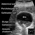
Right upper quadrant abdominal ultrasound is most commonly used to diagnose cholecystitis.[1][26][27] Ultrasound findings suggestive of acute cholecystitis include gallstones, pericholecystic fluid (fluid surrounding the gallbladder), gallbladder wall thickening (wall thickness over 3 mm),[28] dilation of the bile duct, and sonographic Murphy's sign.[13] Given its higher sensitivity, hepatic iminodiacetic acid (HIDA) scan can be used if ultrasound is not diagnostic.[13][14] CT scan may also be used if complications such as perforation or gangrene are suspected.[14]
-
 Abdominal ultrasonography showing gallstones, wall thickening and fluid around the gall bladder
 -
 Gallstones and biliary sludge, but the gallbladder wall is not clearly thickened, with no edema in the pericholecystic fat, thus not cholecystitis.
 -
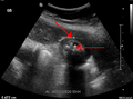 Acute cholecystitis as seen on ultrasound. The closed arrow points to gallbladder wall thickening. Open arrow points to stones in the GB
 -
 Acute cholecystitis with gallbladder wall thickening, a large gallstone, and a large gallbladder
 -
Significant gallbladder wall thickening[29]
-
Significant gallbladder wall thickening[29]


Histopathology
-
 Gross examinationof gallbladder carcinoma, with a prominent nodule.
 -
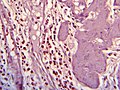
 Histopathology of gallbladder carcinoma, with marked nuclear pleomorphism.
 -
 Histopathology of eosinophilic cholecystitis
 -
 Gross examination of gallbladder cholesterolosis, with yellow streaks of cholesterol deposition.
 -
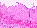 Histopathology of acute gangrenous cholecystitis, showing necrosis, neutrophils and partially sloughed off mucosa.


Differential diagnosis
Many other diagnoses can have similar symptoms as cholecystitis. Additionally the symptoms of chronic cholecystitis are commonly vague and can be mistaken for other diseases. These alternative diagnoses include but are not limited to:[14]
- Perforated peptic ulcer
- Acute pancreatitis
- Liver abscess
- Pneumonia
- Myocardial ischemia
- Hiatal hernia
- Biliary colic
- Choledocholithiasis
- Cholangitis
- Appendicitis
- Colitis
- Acute peptic ulcer exacerbation
- Amoebic liver abscess
- Acute intestinal obstruction
- Kidney stone
- Biliary ascariasis[31]
Treatment

Surgery
For most people with acute cholecystitis, the treatment of choice is surgical removal of the gallbladder, laparoscopic cholecystectomy.[32] Laparoscopic cholecystectomy is performed using several small incisions located at various points across the abdomen. Several studies have demonstrated the superiority of laparoscopic cholecystectomy when compared to open cholecystectomy (using a large incision in the right upper abdomen under the rib cage). People undergoing laparoscopic surgery report less incisional pain postoperatively as well as having fewer long-term complications and less disability following the surgery.[33][34] Additionally, laparoscopic surgery is associated with a lower rate of surgical site infection.[35]
During the days prior to laparoscopic surgery, studies showed that outcomes were better following early removal of the gallbladder, preferably within the first week.[36] Early laparoscopic cholecystectomy (within 7 days of visiting a doctor with symptoms) as compared to delayed treatment (more than 6 weeks) may result in shorter hospital stays and a decreased risk of requiring an emergency procedure.[37] There is no difference in terms of negative outcomes including bile duct injury or conversion to open cholecystectomy.[37] For early cholecystectomy, the most common reason for conversion to open surgery is inflammation that hides Calot's triangle. For delayed surgery, the most common reason was fibrotic adhesions.[37]
Other
Supportive measures may be instituted prior to surgery. These measures include fluid resuscitation. Intravenous
In cases of severe inflammation, shock, or if the person has higher risk for general anesthesia (required for cholecystectomy), an interventional radiologist may insert a percutaneous drainage catheter into the gallbladder (percutaneous cholecystostomy tube) and treat the person with antibiotics until the acute inflammation resolves. A cholecystectomy may then be warranted if the person's condition improves.[41]
Epidemiology
Cholecystitis accounts for 3–10% of cases of abdominal pain worldwide.[43] Cholecystitis caused an estimated 651,829 emergency department visits and 389,180 hospital admissions in the US in 2012.[44] The 2012 US mortality rate was 0.7 per 100,000 people.[44] The frequency of cholecystitis is highest in people age 50–69 years old.[43]
References
- ^ PMID 18579815.
- ^ ISBN 9781437727678. Archivedfrom the original on 2017-09-08.
- ISBN 9780199392148. Archivedfrom the original on 2017-09-08.
- ^ a b c d e f g "Gallstones". NIDDK. November 2013. Archived from the original on 28 July 2016. Retrieved 27 July 2016.
- ^ PMID 27307785.
- ISBN 978-0323076999.
- ^ PMID 26483868.
- ^ )
- ^ ISBN 9781455749898. Archivedfrom the original on 2017-09-08.
- PMID 26468309.
- S2CID 205507793.
- ISBN 9781469835785. Archivedfrom the original on 2017-09-08.
- ^ a b c d e f g h i j k l m n o p q r s t u v w x y z aa ab ac ad Greenberger N.J., Paumgartner G (2012). Chapter 311. Diseases of the Gallbladder and Bile Ducts. In Longo D.L., Fauci A.S., Kasper D.L., Hauser S.L., Jameson J, Loscalzo J (Eds), Harrison's Principles of Internal Medicine, 18e
- ^ a b c d e f g h i j k l m Friedman L.S. (2015). Liver, Biliary Tract, & Pancreas Disorders. In Papadakis M.A., McPhee S.J., Rabow M.W. (Eds), Current Medical Diagnosis & Treatment 2015
- PMID 8780468.
- S2CID 8792764.
- ^ a b "Cholecystitis". Mayo Clinic. 28 August 2014. Archived from the original on 25 November 2014. Retrieved 13 November 2014.
- ^ Barie PS. Acalculous and postoperative cholecystitis. In: Surgical intensive care, Barie PS, Shires GT. (Eds), Little Brown & Co, Boston 1993. p.837.
- PMID 2322038.
- ^ ISBN 978-0-07-183120-8.
- ^ PMID 19653352.
- PMID 15974235.
- PMID 1063276.
- ^ "Boas' sign" (web). GPnotebook. Archived from the original on 2007-02-07. Retrieved 2007-01-21.
- PMID 21250265. Retrieved 27 June 2021 – via National Center for Biotechnology Information, U.S. National Library of Medicine.
- PMID 7979854.
- PMID 3893388.
- PMID 17242260.
- ^ a b "UOTW #30 - Ultrasound of the Week". Ultrasound of the Week. 23 December 2014. Archived from the original on 9 May 2017. Retrieved 27 May 2017.
- PMID 27123469.
- S2CID 19385620.
- PMID 18579815.
- S2CID 26868091.
- PMID 15973772.
- PMID 12616119.
- S2CID 875668.
- ^ PMID 23813477.
- PMID 30656081.
- ^ "Cholecystitis". The Lecturio Medical Concept Library. Retrieved 8 July 2021.
- S2CID 205507793.
- ^ "Acute Cholangitis". The Lecturio Medical Concept Library. Retrieved 27 June 2021.
- ISBN 978-94-010-8181-8.
- ^ PMID 17252293.
- ^ PMID 26327134.