
Chorda tympani
| Chorda tympani | |
|---|---|
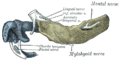
 The left tympanic membrane with the malleus and the chorda tympani, viewed from within the tympanic cavity (medial). | |
| Details | |
| From | Facial nerve |
| Innervates | Taste (anterior 2/3 of tongue) Sublingual gland |
| Identifiers | |
| Latin | nervus chorda tympani |
| MeSH | D002814 |
| TA98 | A14.2.01.084 A14.2.01.118 |
| TA2 | 6292 |
| FMA | 53228 |
| Anatomical terms of neuroanatomy | |
Chorda tympani is a branch of the
Chorda tympani has a complex course from the brainstem, through the temporal bone and middle ear, into the infratemporal fossa, and ending in the oral cavity.[2]
Structure
Chorda tympani fibers emerge from the
Chorda tympani then exits the skull by descending through the petrotympanic fissure into the infratemporal fossa. Here it joins the lingual nerve, a branch of the mandibular nerve (CN V3). Traveling with the lingual nerve, the fibers of chorda tympani enter the sublingual space to reach the anterior 2/3 of the tongue and submandibular ganglion.[4]
- The anterior 2/3 of the tongue and carry taste information to the nucleus of solitary tract of the brainstem, where taste information from facial, glossopharyngeal, and vagusnerves is integrated.
- The superior salivary nucleus of the brainstem and project to the submandibular ganglion to synapse with postganglionic fibers which go on to innervate the submandibular and sublingual salivary glands.[2]
Function
The chorda tympani carries two types of nerve fibers from their origin from the facial nerve to the lingual nerve that carries them to their destinations:
- Special sensory fibers providing taste sensation from the anterior two-thirds of the tongue.
- Preganglionic parasympathetic fibers to the submandibular ganglion, providing secretomotor innervation to two salivary glands: the submandibular gland and sublingual gland and to the vessels of the tongue, which when stimulated, cause a dilation of blood vessels of the tongue.

Taste
The chorda tympani is one of three cranial nerves that are involved in taste. The taste system involves a complicated feedback loop, with each nerve acting to inhibit the signals of other nerves.
There are similarities between the tastes the chorda tympani picks up in sweeteners between mice and
Chorda tympani transection
The chorda tympani nerve carries its information to the nucleus of solitary tract, and shares this area with the greater petrosal, glossopharyngeal, and vagus nerves.[8] When the greater petrosal and glossopharyngeal nerves are cut, regardless of age, the chorda tympani nerve takes over the space in the terminal field. This takeover of space by the chorda tympani is believed to be the nerve reverting to its original state before competition and pruning.[9] The chorda tympani, as part of the
In a bilateral transection of the chorda tympani in mice, the preference for sodium chloride increases compared to before the transection. Also avoidance of higher concentrations of sodium chloride is eliminated.[5] The amiloride-sensitive channels responsible for salt recognition and response is functional in adult rats but not neonatal rats. This explains part of the change in preference of sodium chloride after a chorda tympani transection.[6] The chorda tympani innervates theDysfunction
This section needs expansion. You can help by adding to it. (March 2018) |
Injury to the chorda tympani nerve leads to loss or distortion of taste from anterior 2/3 of tongue.[13] However, taste from the posterior 1/3 of tongue (supplied by the glossopharyngeal nerve) remains intact.
The chorda tympani appears to exert a particularly strong inhibitory influence on other taste nerves, as well as on pain fibers in the tongue. When the chorda tympani is damaged, its inhibitory function is disrupted, leading to less inhibited activity in the other nerves.[citation needed]
Additional images
This gallery of anatomic features needs cleanup to abide by the medical manual of style. ; please improve or remove the gallery accordingly. (May 2015) |
-
 Mandible of human embryo 24 mm. long. Outer aspect.
 -
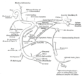
 Distribution of the maxillary and mandibular nerves, and the submaxillary ganglion.
 -
 Plan of the facial and intermediate nerves and their communication with other nerves.
 -
 The course and connections of the facial nerve in the temporal bone.
 -
 Sympathetic connections of the submaxillary and superior cervical ganglia.
 -
 View of the inner wall of the tympanum (enlarged.)
 -
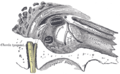
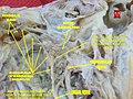 Dissection of chorda tympani nerve
 -
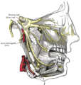
 Lateral head anatomy detail.Facial nerve dissection.


References
External links
- Anatomy figure: 27:03-08 at Human Anatomy Online, SUNY Downstate Medical Center
- "7-18". Cranial Nerves. Yale School of Medicine. Archived from the original on 2016-03-03.
- MedEd at Loyola GrossAnatomy/h_n/cn/cn1/cnb7c.htm
- cranialnerves at The Anatomy Lesson by Wesley Norman (Georgetown University) (VII)
- Photo at Washington University in St. Louis