
Choroid
| Choroid | |
|---|---|
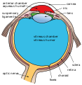
 Cross-section of human eye, with choroid labeled at top. | |
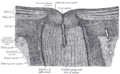
 Interior of anterior half of bulb of eye. (Choroid labeled at right, second from the bottom.) | |
| Details | |
| Artery | short posterior ciliary arteries, long posterior ciliary arteries |
| Identifiers | |
| Latin | choroidea |
| MeSH | D002829 |
| TA98 | A15.2.03.002 |
| TA2 | 6774 |
| FMA | 58298 |
| Anatomical terminology | |
The choroid, also known as the choroidea or choroid coat, is a part of the uvea, the vascular layer of the eye. It contains connective tissues, and lies between the retina and the sclera. The human choroid is thickest at the far extreme rear of the eye (at 0.2 mm), while in the outlying areas it narrows to 0.1 mm.[1] The choroid provides oxygen and nourishment to the outer layers of the retina. Along with the ciliary body and iris, the choroid forms the uveal tract.
The structure of the choroid is generally divided into four layers (classified in order of furthest away from the retina to closest):
- Haller's layer - outermost layer of the choroid consisting of larger diameter blood vessels;[1]
- Sattler's layer - layer of medium diameter blood vessels;[1]
- Choriocapillaris - layer of capillaries;[1] and
- Bruch's membrane (synonyms: Lamina basalis, Complexus basalis, Lamina vitra) - innermost layer of the choroid.[1]
Blood supply
There are two circulations of the eye: the retinal (in the retina) and

In bony fish
Mechanism
Melanin, a dark colored pigment, helps the choroid limit uncontrolled reflection within the eye that would potentially result in the perception of confusing images.
In humans and most other
History
The choroid was first described by
About 100 years later, Herophilos (c. 335 – 280 BCE) also described the choroid from his dissections on eyes of cadavers.[10][11]
Clinical significance
Choroid is the most common site for metastasis in the eye due to its extensive vascular supply. The origin of the metastases are usually from breast cancer, lung cancer, gastrointestinal cancer, and kidney cancer. Bilateral choroidal metastases are usually due to breast cancer, while unilateral metastasis is due to lung cancer. Choroidal metastases should be differentiated from uveal melanoma, where the latter is a primary tumour arising from the choroid itself.[12]
See also
Additional images
-
 Schematic cross section of the human eye; choroid is shown in purple.
 -
 Laser Doppler imaging of retinal and choroidal blood flow
 -
 Iris, front view
 -
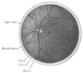
 The terminal portion of the optic nerve and its entrance into the eyeball, in horizontal section
 -
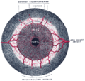
 The interior of the posterior half of the left eyeball
 -
 Structures of the eye labeled
 -
 This image shows another labeled view of the structures of the eye
 -
 Calf's eye dissected to expose the choroid: its tapetum lucidum is iridescent blue


References
- ^ a b c d e MRCOphth Sacs questions
- ^ The Ocular Circulation
- ^ "Sensory Reception: Human Vision: Structure and function of the Human Eye" vol. 27, p. 174 Encyclopædia Britannica, 1987
- PMID 812547.
- ^ Puyo, Léo, Michel Paques, Mathias Fink, José-Alain Sahel, and Michael Atlan. "Choroidal vasculature imaging with laser Doppler holography." Biomedical optics express 10, no. 2 (2019): 995-1012.
- ^ Léo Puyo, Michel Paques, and Michael Atlan, "Spatio-temporal filtering in laser Doppler holography for retinal blood flow imaging," Biomed. Opt. Express 11, 3274-3287 (2020)
- ^ "Eye (Vertebrate)" McGraw-Hill Encyclopedia of Science and Technology, vol. 6, 2007.
- ^ Dolz-Marco, R., Gallego-Pinazo, R., Dansingani, K. K., & Yannuzzi, L. A. (2017). The history of the choroid. In J. Chhablani & J. Ruiz-Medrano (Eds.), Choroidal Disorders (Vol. 1–5, pp. 1-5). Academic Press. https://doi.org/0.1016/b978-0-12-805313-3.00001-6
- ISSN 0009-8388.
- ISBN 978-0-521-23646-1.
- ^ Reverón, R. (2015). Herophilos, the great anatomist of antiquity. Anatomy, 9(2), 108-111. https://doi.org/10.2399/ana.15.003 https://dergipark.org.tr/en/download/article-file/371071
- PMID 25827542.
External links
- Histology image: 08008loa – Histology Learning System at Boston University
| Fibrous tunic (outer) |
|  | |||||
|---|---|---|---|---|---|---|---|
| Uvea / vascular tunic (middle) |
| ||||||
| Retina (inner) |
| ||||||
| Anatomical regions of the eye |
| ||||||
| Other |
| ||||||