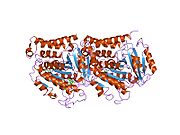
Class III β-tubulin
| TUBB3 | |||
|---|---|---|---|
Gene ontology | |||
| Molecular function | |||
| Cellular component | |||
| Biological process | |||
| Sources:Amigo / QuickGO | |||
Ensembl | |||||||||
|---|---|---|---|---|---|---|---|---|---|
| UniProt | |||||||||
| RefSeq (mRNA) | |||||||||
| RefSeq (protein) | |||||||||
| Location (UCSC) | Chr 16: 89.92 – 89.94 Mb | Chr 8: 124.14 – 124.15 Mb | |||||||
| PubMed search | [3] | [4] | |||||||
| View/Edit Human | View/Edit Mouse |
Class III β-tubulin, otherwise known as βIII-tubulin (β3-tubulin) or β-tubulin III, is a
It is possible to use
Class III β-tubulin is one of the seven
Gene
The human TUBB3 gene is located on chromosome 16q24.3, and consists of 4 exons that transcribe a protein of 450aa. A shorter isoform of 378aa derived from alternative splicing of exon 1 is devoid of part of the N-terminus and may be responsible for mitochondrial expression.[12][20] Like other β-tubulin isotypes, βIII-tubulin has a GTPase domain which plays an essential role in regulating microtubule dynamics.[21] Differences between Class I (the most commonly represented and constitutively expressed isotype) and class III β-tubulin are limited to only 13aa within region 1-429aa, while all amino acids in region 430-450aa are divergent. These variations in primary structure affect the paclitaxel (a mimic of Nur77) binding domain on βIII-tubulin and may account for the ability of this isotype to confer resistance to Nur77-initiated apoptosis.[22]
Function
Cysteine residues in class III β-tubulin are actively involved in regulating ligand interactions and microtubule formation. Proteomic analysis has revealed that many factors bound to these cysteine residues are involved in the oxidative stress and glucose deprivation response.[12] This is particularly interesting in light of the fact that class III β-tubulin first appears in the phylogenetic tree when life emerged from the seas and cells were exposed to atmospheric oxygen.[23] In structural terms, constitutive Class I (TUBB) and Class IVb (TUBB2C) β-tubulins contain a cysteine at position 239, while βIII-tubulin has a cysteine at position 124. Position 239 can be readily oxidized while position 124 is relatively resistant to oxidation.[24] Thus, a relative abundance of βIII-tubulin in situations of oxidative stress could provide a protective benefit.
Interactions
The interactome of class III β-tubulin comprises the GTPase GBP1 (guanylate binding protein 1) and a panel of an additional 19 kinases having prosurvival activity including PIM1 (Proviral Integration Site 1) and NEK6 (NIMA-related kinase 6). Incorporation of these kinases into the cytoskeleton via the GBP-1/ class III β-tubulin interaction protects kinases from rapid degradation.[25] Other pro-survival factors interacting with class III β-tubulin enabling cellular adaptation to oxidative stress include the molecular chaperone HSP70/GRP75.[26] FMO4 (vimentin/dimethylalanine monooxygenase 4) and GSTM4 (glutathione transferase M4).[12]
Regulation
The expression of Class III β-tubulin is regulated at both the transcriptional and translational levels. In neural tissue, constitutive expression is driven by Sox4 and Sox11.[27] In non-neural tissues, regulation is dependent on an E-box site in the 3' flanking region at +168 nucleotides. This site binds basic helix-loop-helix (bHLH) hypoxia induced transcription factors Hif-1α and Hif-2α and is epigenetically modified in cancer cells with constitutive TUBB3 expression.[14][28] Translational regulation of TUBB3 occurs in the 3`flanking region with the interaction of the miR-200c family of micro-RNA.[29][30] MiR-200c is in turn modulated by the protein HuR (encoded by ELAVL1). When HuR is predominantly in the nucleus, a phenomenon typically occurring in low stage carcinomas, miR-200c suppresses class III β-tubulin translation. By contrast, cytoplasmic HuR and miR-200c enhance class III β-tubulin translation by facilitating the entry of the mRNA into the ribosome.[15][31]
Role in cancer
In oncology, class III β-tubulin has been investigated as both a prognostic biomarker and an indicator of resistance to taxanes and other compounds.[32][33] The majority of reports implicate class III β-tubulin as a biomarker of poor outcome. However, there are also data in clear cell carcinoma, melanoma and breast cancer showing favorable prognosis.[34][35][36][37] Class III β-tubulin is integral component of a pro-survival, cascading molecular pathway which renders cancer cells resistant to apoptosis and enhances their ability to invade local tissues and metastasize.[14][38][39][40] Class III β-tubulin performs best as a prognostic biomarker when analyzed in the context of an integrated signature including upstream regulators and downstream effectors.[15][31][41] TUBB3 mutation is associated with microlissencephaly.
Overexpression of this isotype in clinical samples correlates with tumor aggressiveness, resistance to chemotherapeutic drugs, and poor patient survival.[42][43]
Pathophysiology
The β3 isotype increases tumor aggressiveness by two distinct mechanisms. Incorporation of this isotype makes microtubule networks hypostable, allowing them to resist the cytotoxic effects of microtubule stabilizing drugs like taxanes or epothilones. Mechanistically, it was found that overexpression of β3-tubulin increases the rate of microtubule detachment from microtubule organizing centers, an activity that is suppressed by drugs such as paclitaxel.[44]
Expression of β3-tubulin also makes cells more aggressive by altering their response to drug-induced suppression of microtubule dynamics.[45] Dynamic microtubules are needed for the cell migration that underlies processes such as tumor metastasis and angiogenesis. The dynamics are normally suppressed by low, subtoxic concentrations of microtubule drugs that also inhibit cell migration. However, incorporating β3-tubulin into microtubules increases the concentration of drug that is needed to suppress dynamics and inhibit cell migration. Thus, tumors that express β3-tubulin are not only resistant to the cytotoxic effects of microtubule targeted drugs, but also to their ability to suppress tumor metastasis. Moreover, expression of β3-tubulin also counteracts the ability of these drugs to inhibit angiogenesis which is normally another important facet of their action.
Notes
PMID 25839941 . |
References
- ^ a b c GRCh38: Ensembl release 89: ENSG00000258947 – Ensembl, May 2017
- ^ a b c GRCm38: Ensembl release 89: ENSMUSG00000062380 – Ensembl, May 2017
- ^ "Human PubMed Reference:". National Center for Biotechnology Information, U.S. National Library of Medicine.
- ^ "Mouse PubMed Reference:". National Center for Biotechnology Information, U.S. National Library of Medicine.
- PMID 3459176.
- PMID 2817080.
- ^ PMID 18220531.
- PMID 21734264.
- PMID 9473684.
- PMID 8098743.
- ^ "Entrez Gene: TUBB3 tubulin, beta 3".
- ^ S2CID 26212762.
- ^ PMID 20829227.
- ^ PMID 18178340.
- ^ PMID 20587520.
- PMID 11300931.
- S2CID 10388161.
- PMID 24179174.
- PMID 20074521.
- PMID 11034069.
- PMID 22390762.
- PMID 19671798.
- PMID 16479502.
- PMID 18435451.
- S2CID 24222768.
- S2CID 16958013.
- PMID 17182872.
- PMID 19074887.
- PMID 19435871.
- PMID 20049172.
- ^ PMID 23394580.
- PMID 21999149.
- S2CID 26229777.
- S2CID 25857848.
- S2CID 7703873.
- PMID 23218766.
- PMID 19122647.
- PMID 17909044.
- PMID 20501838.
- PMID 25414139.
- PMID 24661907.
- S2CID 26229777.
- S2CID 85980035.
- PMID 21741453.
- PMID 21576762.


