
Conjunctiva
| Conjunctiva | |
|---|---|
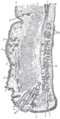
 The upper half of a sagittal section through the front of the eyeball. (Label for 'Conjunctiva' visible at center-left) | |
 Horizontal section of the eyeball. (Conjunctiva labeled at upper left) | |
| Details | |
| Part of | Eye |
| Artery | lacrimal artery, anterior ciliary arteries |
| Nerve | supratrochlear nerve |
| Identifiers | |
| Latin | tunica conjunctiva |
| MeSH | D003228 |
| TA98 | A15.2.07.047 |
| TA2 | 6836 |
| FMA | 59011 |
| Anatomical terminology | |


In the
Structure
The conjunctiva is typically divided into three parts:
| Part | Area |
|---|---|
| Palpebral or tarsal conjunctiva | Lines the eyelids |
| Bulbar or ocular conjunctiva | Covers the eyeball, over the anterior sclera: This region of the conjunctiva is tightly bound to the underlying sclera by Tenon's capsule and moves with the eyeball movements. The average thickness of the bulbar conjunctival membrane is 33 microns.[2] |
| Fornix conjunctiva | Forms the junction between the bulbar and palpebral conjunctivas: It is loose and flexible, allowing the free movement of the lids and eyeball.[3] |
Blood supply
Blood to the bulbar conjunctiva is primarily derived from the ophthalmic artery. The blood supply to the palpebral conjunctiva (the eyelid) is derived from the external carotid artery. However, the circulations of the bulbar conjunctiva and palpebral conjunctiva are linked, so both bulbar conjunctival and palpebral conjunctival vessels are supplied by both the ophthalmic artery and the external carotid artery, to varying extents.[4]
Nerve supply
Sensory innervation of the conjunctiva is divided into four parts:[5]
| Area | Nerve |
|---|---|
| Superior | |
| Inferior | Infraorbital nerve |
| Lateral | Lacrimal nerve (with contribution from zygomaticofacial nerve) |
| Circumcorneal | Long ciliary nerves |
Microanatomy
The conjunctiva consists of unkeratinized, both stratified squamous and stratified columnar epithelium, with interspersed
Function
The conjunctiva helps lubricate the eye by producing mucus and tears, although a smaller volume of tears than the lacrimal gland.[7] It also contributes to
Clinical significance
Disorders of the conjunctiva and
- The conjunctival microvascular hemodynamics are affected by diabetic retinopathy (DR), hence can be useful for DR diagnosis and monitoring,[8] and discriminating stages of DR.[9]
- Hypertension is associated with an increase in the tortuosity of bulbar conjunctival blood vessels and capillary and arteriole loss.[17][18]
- Carotid artery occlusion is associated with slower conjunctival blood flow and apparent capillary loss.[4]
- With age, the conjunctiva can stretch and loosen from the underlying sclera, leading to the formation of conjunctival folds, a condition known as conjunctivochalasis.[19][20]
- The conjunctiva can be affected by tumors which can be benign, pre-malignant or malignant.[21]
- Leptospirosis, an infection with Leptospira, can cause conjunctival suffusion, which is characterized by chemosis, and redness without exudates.
Bulbar conjunctival microvasculature
Vessel morphology
The bulbar conjunctival
Blood oxygen dynamics
The bulbar conjunctival microvasculature is in close proximity to ambient air, thus
Blood vessel imaging methods
The bulbar conjunctival microvessels are typically imaged with a high-magnification slit lamp with green filters.[25][26][27] With such high-magnification imaging systems, it is possible to see groups of individual red blood cells flowing in vivo.[25] Fundus cameras may also be used for low-magnification wide field-of-view imaging of the bulbar conjunctival microvasculature. Modified fundus cameras have been used to measure conjunctival blood flow [28] and to measure blood oxygen saturation.[24] Fluorescein angiography has been used to study the blood flow of the bulbar conjunctiva and to differentiate the bulbar conjunctival and episcleral microcirculation.[29][30][31]
Vasodilation
The bulbar conjunctival microvasculature is known to dilate in response to several stimuli and external conditions, including allergens (e.g. pollen),[32] temperature,[33] time-of-day,[33] contact-lens wear,[13] and acute mild hypoxia.[24] Bulbar conjunctival vasodilation has also been shown to correlate changes in emotional state.[34]
Type 2 diabetes is associated with an increase in average bulbar conjunctival vessel diameter and capillary loss.
See also
- Conjunctivitis (pink-eye)
- Conjunctivochalasis
- Dry eye
- Pinguecula
- Pterygium
- Rougine
- Subconjunctival hemorrhage
- Diabetes
- Sickle-cell disease
- Slit lamp
Additional images
-
 Sagittal section through the upper eyelid.
 -
 Extrinsic eye muscle. Nerves of orbita. Deep dissection.


References
- ^ "Conjunctiva". www.sciencedirect.com. Retrieved 4 August 2022.
- S2CID 35398240.
- ^ Eye, human Encyclopædia Britannica
- ^ PMID 13660526.
- ^ "Table 1: Summary of sensory nerve supply". Archived from the original on February 14, 2013. Retrieved July 31, 2016.
- ^ ISBN 978-1437727883.
- ^ London Place Eye Center (2003). Conjunctivitis Archived 2004-08-08 at the Wayback Machine. Retrieved July 25, 2004.
- PMID 28387229.
- PMID 27446692.
- PMID 3759367.
- ^ PMID 491983.
- ^ PMID 5236035.
- ^ PMID 11585371.
- ^ PMID 5720854.
- PMID 3800533.
- PMID 23657867.
- PMID 748720.
- S2CID 11589129.
- ^ "Conjunctivochalasis - Medical Definition". Medilexicon.com. Archived from the original on 2016-03-03. Retrieved 2012-11-13.
- .
- PMID 21234216.
- PMID 13364178.
- PMID 20053367.
- ^ S2CID 25038785.
- ^ S2CID 1595902.
- PMID 27446692.
- PMID 26452274.
- PMID 23084966.
- PMID 3256492.
- PMID 8543081.
- PMID 3814565.
- PMID 8828525.
- ^ S2CID 943038.
- ISSN 1439-0310.
External links
- "Conjunctiva". Medicinenet.com. 1999. Archived from the original on 7 June 2014. Retrieved 25 July 2004.