
Coronary sinus
This article includes a improve this article by introducing more precise citations. (June 2015) ) |
| Coronary sinus | |
|---|---|
Right atrium | |
| Identifiers | |
| Latin | sinus coronarius |
| MeSH | D054326 |
| TA98 | A12.3.01.002 |
| TA2 | 4158 |
| FMA | 4706 |
| Anatomical terminology] | |
The coronary sinus (from
Structure
Origin
The coronary sinus arises upon the posterior aspect of the heart between the
Course
The coronary sinus runs transversely in the left
Fate
The coronary sinus drains through the posterior wall of right atrium at the orifice of the coronary sinus.
Tributaries
The coronary sinus receives blood mainly from the
- Great cardiac vein (run upwards in the anterior interventricular sulcus to the left atrioventricular groove to form the coronary sinus;[6]
- Middle cardiac vein (ascends posterior interventricular sulcus to drain into coronary sinus);[6]
- Small cardiac vein (accompanies right coronary artery in the right atrioventricular groove to drain into the right side of the coronary sinus;[6]
- Posterior vein of left ventricle (accompanies the left marginal artery, ascends the posterior wall of left ventricle to drain into the coronary sinus);[6]
- Oblique vein of left atrium.[1]
All veins that empty into the coronary sinus except for the oblique vein of the left atrium have valves at their junction with the coronary sinus.[1]
The
Microanatomy
The wall of the coronary sinus is partly muscular.[citation needed]
Function
The coronary sinus is responsible for venous return of about 55% of the cardiac blood supply.[1]
Clinical significance
Electrodes can be inserted into and through the coronary sinus to study the electrophysiology of the heart. This includes for a coronary sinus electrogram.[5] The coronary sinus connects directly with the right atrium. It will dilate as a result of any condition that causes elevated right atrial pressure, such as pulmonary hypertension.[7] Dilated coronary sinus is also seen in some congenital cardiovascular conditions, such as persistent left superior vena cava,[8] and total anomalous pulmonary venous return.[9]
Additional images
-
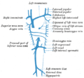 Diagram showing completion of development of the parietal veins.
 -
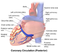 Posterior view of coronary circulation


See also
References
- ^ PMID 32491498, retrieved 2023-01-05
- ^ OCLC 1044772257.)
{{cite book}}: CS1 maint: location missing publisher (link - ^ PMID 22786990.
- ISBN 9783642659836.
- ^ ISBN 978-0-323-52356-1, retrieved 2021-01-12
- ^ ISBN 9780702029714.
- PMID 11174433.
- PMC PMC7157846.
- PMID 33904266
External links
- Anatomy figure: 20:04-03 at Human Anatomy Online, SUNY Downstate Medical Center – "Posterior view of the heart."
- MedEd at Loyola Radio/curriculum/Vascular/Coronary_sinus.jpg