Coronavirus envelope protein

Source: Wikipedia, the free encyclopedia.
Envelope protein
virion[1]
Blue: envelope
Turquoise: spike glycoprotein (S)
Bright Pink: envelope proteins (E)
Green: membrane proteins (M)
Orange: glycans
Identifiers
SymbolCoV_E
PfamPF02723
InterProIPR003873
PROSITEPS51926
Available protein structures:
Pfam  structures / ECOD  
PDBRCSB PDB; PDBe; PDBj
PDBsumstructure summary

The envelope (E) protein is the smallest and least well-characterized of the four major

viral capsid, it is thought to be involved in viral assembly, likely functions as a viroporin, and is involved in viral pathogenesis.[2][5]

Structure

cations.[5][4] Rendered from PDB: 7K3G
​.

The E protein consists of a short

conserved only in the alpha and beta coronavirus groups, but not gamma.[2] In the beta and gamma groups, a conserved proline residue is found in the C-terminal region likely involved in targeting the protein to the Golgi.[2]

The transmembrane helices of the E proteins of SARS-CoV and SARS-CoV-2 can

cation-selective ion channels.[5] Both viruses' E protein pentamers have been structurally characterized by nuclear magnetic resonance spectroscopy.[5][7]

The membrane topology of the E protein has been studied in a number of coronaviruses with inconsistent results; the protein's orientation in the membrane may be variable.[3] The balance of evidence suggests the most common orientation has the C-terminus oriented toward the cytoplasm.[8] Studies of SARS-CoV-2 E protein are consistent with this orientation.[5][9]

Post-translational modifications

In some, but not all, coronaviruses, the E protein is

Ubiquitination of SARS-CoV E has also been described, though its functional significance is also not known.[2]

Expression and localization

Genomic information
Genomic organisation of isolate Wuhan-Hu-1, the earliest sequenced sample of SARS-CoV-2, indicating the location of the E gene
NCBI genome ID86693
Genome size29,903 bases
Year of completion2020
Genome browser (UCSC)

The E protein is

virions.[2][4] E protein is localized to the endoplasmic reticulum, Golgi apparatus, and endoplasmic-reticulum–Golgi intermediate compartment (ERGIC), the intracellular compartment that gives rise to the coronavirus viral envelope.[2][5]

Function

Essentiality

Studies in different coronaviruses have reached different conclusions about whether E is

viral titer,[12] in some cases by introducing propagation defects or causing abnormal capsid morphology.[2]

Virions and viral assembly

respiratory mucosa, showing the positions of the four structural proteins and components of the extracellular environment[13]

The E protein is found in assembled virions where it forms

viral budding or scission, although its role in this process has not been well characterized.[2][4][15]

Viroporin

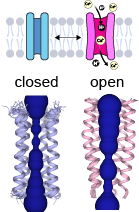
The E viroporin opens at acid pH. The open state in pink presents a wide N-terminus. Conversely, the C-terminus narrows in the open state, which brings the polar sidechains of Thr35 and Arg38 close to the hydrophobic gate at Leu28. This presumedly lowers the energy barrier for ions to cross the channel.

In its

cation-selective ion channels and likely functions as a viroporin.[5] NMR studies show that viroporin presents an open conformation at low pH or in the presence of calcium ions, while the closed conformation is favored at basic pH.[16] The NMR structure shows a hydrophobic gate at leucine 28 in the middle of the pore. The passage of ions through the gate is thought to be facilitated by the polar residues at the C-terminus.[17]

The cation leakage may disrupt ion

membrane permeability, and modulate pH in the host cell, which may facilitate viral release.[2][4]


The E protein's role as a viroporin appears to be involved in

animal models despite little effect on viral growth.[10]


Interactions with host proteins

Cryo-electron microscopy structure of the interaction between the SARS-CoV-2 E protein PDZ-binding motif (magenta) and a construct containing the PDZ (blue), SH3 (yellow), and guanylate kinase-like (GK, green) domains from a host cell protein, human PALS1[19]

SARS-CoV and occur via the C-terminal PDZ domain binding motif. The SARS-CoV E protein has been reported to interact with five host cell proteins: Bcl-xL, PALS1, syntenin, sodium/potassium (Na+/K+) ATPase α-1 subunit, and stomatin.[2] The interaction with PALS1 may be related to pathogenesis via the resulting disruption in tight junctions.[3][10] This interaction has also been identified in SARS-CoV-2.[19]

Evolution and conservation

The sequence of the E protein is not well

deletion.[4] A study of SARS-CoV-2 sequences suggests that the E protein is evolving relatively slowly compared to other structural proteins.[21] The conserved nature of the envelope protein among SARS-CoV and SARS-CoV-2 variants has led it to be researched as a potential target for universal coronavirus vaccine development.[22][23]

References

  1. ^ Solodovnikov, Alexey; Arkhipova, Valeria (2021-07-29). "Достоверно красиво: как мы сделали 3D-модель SARS-CoV-2" [Truly beautiful: how we made the SARS-CoV-2 3D model] (in Russian). N+1. Archived from the original on 2021-07-30. Retrieved 30 July 2021.
  2. ^
    PMID 31133031
    .
  3. ^ .
  4. ^ .
  5. ^ .
  6. .
  7. .
  8. ^ .
  9. .
  10. ^ .
  11. .
  12. ^ .
  13. .
  14. .
  15. ^ .
  16. .
  17. .
  18. .
  19. ^ .
  20. .
  21. .
  22. .
  23. .