Cranioplasty
| Cranioplasty | |
|---|---|
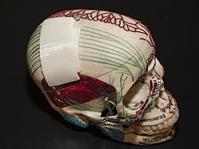
 3D printed prosthesis for cranioplasty | |
| ICD-9-CM | 02.0 |
Cranioplasty is a
Cranioplasty was closely related to trephination and the earliest operation is dated to 3000 BC.[2] Currently, the procedure is performed for both cosmetic and functional purposes. Cranioplasty can restore the normal shape of the skull and prevent other complications caused by a sunken scalp, such as the "syndrome of the trephined".[3] Cranioplasty is a risky operation, with potential risks such as bacterial infection and bone flap resorption.[4]
Etymology
The word cranioplasty can be broken down into two parts: cranio- and -plasty. Cranio- originates from the Ancient Greek word κρανίον, meaning "cranium", while -plasty comes from the Ancient Greek word πλαστός, meaning "moulded" or "fashioned".[5]
Medical uses
The operation has its cosmetic value as the normal shape of the cranium of patients is restored instead of the presence of a sunken skin flap, which may affect the confidence of patients.[1][6]
It also has its therapeutic value as the operation provides structure to the skull and protection to the brain from physical damage.[1][6] The surgery restores regular cerebrospinal fluid (CSF) and cerebral blood flow dynamics, along with normal intracranial pressure.[1][3] Cranioplasty may improve neurological function in some individuals. Furthermore, it can reduce the occurrence of headaches caused by injury or previous surgery.[6]
The optimal timing of cranioplasty is controversial among literature. Some literature stated that the time between a craniectomy and a cranioplasty is usually between 6 months to a year,[1] while others stated that the two operations should be more than a year apart.[7]
The timing of cranioplasty is affected by multiple factors. Sufficient time is required for the recovery of the incision from the previous operation, as well as to clear any infections (both systemic and cranial).[1] Some findings showed that a greater infection rate is associated with early cranioplasty due to interruption of wound healing,[8] as well as an increased incidence of hydrocephalus.[9] Contrarily, there is evidence of early cranioplasty limiting complications caused by "syndrome of the trephined", including changes in cerebral blood flow and abnormal cerebrospinal fluid hydrodynamics.[8] Other researchers reported no significant difference in infection rate with different operational timings.[8][9]
Contraindications are circumstances that indicate the treatment or operation should not be provided due to potential harm. Contraindications for cranioplasty include the presence of bacterial infection, brain swelling, and hydrocephalus.[10] Cranioplasty is withheld until all contraindications are cleared.[citation needed]
Procedure

Before the operation, CT scans and MRIs are taken to study the cranial defect. The patient is given antibiotics to prevent bacterial infection.[1]
The patient is situated on a
After the operation, a
Children
Special considerations to children undergoing cranioplasty are made to accommodate for their growing cranium. Certain materials are more favoured when compared to adult cranioplasty.[citation needed]
Autologous bone grafts are the most preferred materials for paediatric cranioplasty, as they are accepted by the host and the bone flap can be integrated into the body of the host.[10] However, autologous bone pieces may be unavailable or unsuitable in certain occasions. The body size of children may be not enough to have bone flaps to be stored in their subcutaneous spaces, while cryopreservation facilities for bone grafts are not widely available.[11][12] The use of autograft is also associated with a high rate of bone resorption.[11]
Synthetic materials are used for paediatric cranioplasty when the use of autografts is not available or not recommended. Hydroxyapatite is another option for children cranioplasty as it allows the expansion of cranium for children and its ability to be moulded smoothly. It is less commonly used than autografts due to its brittle nature, high infection rate, and poor ability to integrate with the human cranium.[10]
Bilateral cranioplasties are more prone to complications compared to unilateral cranioplasties in children. This may be explained by its larger scalp wound area, a higher volume of blood loss, and the higher complexity and duration of the operation.[12]
Risks
Cranioplasty is an operation with a complication risk ranging from 15 to 41%.[4] The cause for such a high risk of complication compared to other neurosurgical operations is unclear. Male patients and older patients are groups with higher rates of complication.[4]
Complications occurring after cranioplasty include
The risk of bacterial infections in performing cranioplasty ranges from 5 to 12.8%.[1] Multiple factors are affecting the risk of infection, one being the materials used for the operation. Using titanium, whether being custom-made or using a mesh, is associated with a lower infection rate;[1][13] on the other hand, materials such as methyl methacrylate and autologous bone is associated with a higher infection rate.[1] Another risk factor for bacterial infection is the location of the operation. Bifrontal cranioplasties are associated with significantly higher infection rates and higher rates for reoperation.[9][14] Other risk factors for infection include previous infections, contact between sinuses and operation site, devascularized scalp (loss of blood supply in the scalp), previous operations, and type of injury.[1]
Bone resorption is another complication of cranioplasty with a complication rate of 0.7-17.4%.[1] Bone resorption occurs when the autologous graft does not have blood supply due to devitalisation, or when scar tissues or soft tissues remain on the edge of the cranial defect during cranioplasty.[1] Paediatric patients have a higher risk of resorption,[1][4][9] with a resorption rate up to 50%.[4] Bone resorption is more likely to occur in this group of patients when their cranioplasty is carried out over 6 weeks from their previous operation.[9] Fragmented bone flaps, as well as large bone flaps (>70 cm2), are associated with a higher resorption rate.[4]
History
Ancient history

The earliest cranioplasty operation is dated to 3000 BC in the Pre-Columbian Peruvian civilisation, where precious metals, gourds, and shells were found next to trepanned skulls in graveyards, suggesting that cranioplasty had been performed.[2] In the Paracas region of present-day Peru, a skull from 2000 BC with a thin plate of gold covering a cranial defect was found.[2] Moreover, defective skulls were found covered with coconut shells or palm leaves in ancient tribes of the Polynesian Islands.[15] Sanan and Haines stated that the materials being used for cranioplasty were associated with the status of the patient.[2]
Modern history
Research on cranioplasty was not emphasised among early surgical authors in ancient

The earliest modern description of cranioplasty was written by surgeon Ibrahim bin Abdullah of the Ottoman Empire, in his surgical book Alâim-i Cerrâhîn in 1505. The book mentioned the use of xenografts from Kangal dogs or goats as materials for cranioplasty. Such materials were used due to the accessibility of these animals near battlefields, where the procedure is likely to be performed.[16]
The first true description of cranioplasty in Europe was made by
Since the first operation, bones from more animal species were used as xenografts for cranioplasty. These include dogs, apes, geese, rabbits, calves, eagles, oxen, and buffalos. In 1917, William Wayne Babcock reported the use of "soup bone", a piece of cooked and perforated animal bone as a xenograft.[2][15]
Development of modern materials
The prevalence of

Autografts, or autologous grafts, are body tissues taken from the patient. The first successful cranioplasty using an autograft was recorded in 1821, with the bone piece being reinserted into the cranium. The operation achieved partial healing.[2][10] Subsequently, more studies and operations were carried out with autografts. A successful case of reimplantation of cranial bone was reported by Sir William Macewen in 1885, popularising autografts to be material for cranioplasty. Succeeding operations involved autografts taken from different parts of the patient's body, such as the tibia (leg bone), scapula (shoulder blade), ilium (hip bone), sternum (chest bone), along with fat tissues and fascia.[2]
The use of methyl methacrylate (PMMA) for cranioplasty was being developed since World War II, and the material is used extensively since 1954,[2][17][10][18] when there is a high demand for cranioplasty due to a large number of injuries. It becomes malleable when an exothermic reaction occurs between its powder form and benzoyl peroxide, allowing it to be moulded to the cranial defect.[1] Advantages of using PMMA is its malleability, low cost, high strength and high durability. Its disadvantages include being vulnerable to infection as bacteria may adhere to its fibrous layer, as well as its brittle nature and having no growth potential.[1]
Other common synthetic materials for cranioplasty include titanium and hydroxyapatite. Titanium was first used for cranioplasty in 1965.[2] It can be used as a plate, a mesh, and be 3D printed as a porous form.[19] Titanium is non-ferromagnetic and non-corrosive, making the host free from inflammatory reactions.[1] It is also robust, thus preventing patients from trauma.[19] The use of titanium is associated with a lower infection rate.[10] Disadvantages of using titanium include its high cost, poor malleability, and disruption to CT scan images.[1]
Hydroxyapatite is a compound of
See also
- Human cranium
- Craniotomy
- Decompressive craniectomy
- Oral and maxillofacial surgery
- Plastic surgery
- Craniofacial surgery
References
- ^ PMID 28325460.
- ^ PMID 9055300.
- ^ PMID 9068693.
- ^ a b c d e f g Acciarri N, Nicolini F, Martinoni M (December 2016). "Cranioplasty: Routine Surgical Procedure or Risky Operation?". World Journal of Surgical Research. 5 (5).
- ^ "Cranioplasty". Oxford English Dictionary (Online ed.). Oxford University Press. (Subscription or participating institution membership required.)
- ^ a b c "Cranioplasty". Johns Hopkins Medicine. Retrieved 25 May 2020.
- S2CID 2037637.
- ^ S2CID 4505995.
- ^ S2CID 32109527.
- ^ S2CID 22048837.
- ^ PMID 26000090.
- ^ S2CID 7604976.
- PMID 25482456.
- .
- ^ ISBN 978-3-7091-8764-7.
- PMID 17719987.
- ^ PMID 24684330.
- ^ PMID 21897681.
- ^ PMID 32063880.
