
Diatom
| Diatoms Temporal range:
| |
|---|---|

| |
| Light microscopy of a sampling of marine diatoms found living between crystals of annual sea ice in Antarctica, showing a multiplicity of sizes and shapes | |
| Scientific classification | |
| Domain: | Eukaryota |
| Clade: | Diaphoretickes |
| Clade: | SAR |
| Clade: | Stramenopiles |
| Phylum: | Gyrista |
| Subphylum: | Ochrophytina |
| Infraphylum: | Diatomista
|
| Superclass: | Khakista
|
| Class: | Bacillariophyceae Dangeard, 1933[1] |
| Synonyms | |
| |
A diatom (Neo-Latin diatoma)[a] is any member of a large group comprising several genera of algae, specifically microalgae, found in the oceans, waterways and soils of the world. Living diatoms make up a significant portion of the Earth's biomass: they generate about 20 to 50 percent of the oxygen produced on the planet each year,[10][11] take in over 6.7 billion tonnes of silicon each year from the waters in which they live,[12] and constitute nearly half of the organic material found in the oceans. The shells of dead diatoms can reach as much as a half-mile (800 m) deep on the ocean floor, and the entire Amazon basin is fertilized annually by 27 million tons of diatom shell dust transported by transatlantic winds from the African Sahara, much of it from the Bodélé Depression, which was once made up of a system of fresh-water lakes.[13][14]
Diatoms are unicellular organisms: they occur either as solitary cells or in colonies, which can take the shape of ribbons, fans, zigzags, or stars. Individual cells range in size from 2 to 200 micrometers.[15] In the presence of adequate nutrients and sunlight, an assemblage of living diatoms doubles approximately every 24 hours by asexual multiple fission; the maximum life span of individual cells is about six days.[16] Diatoms have two distinct shapes: a few (centric diatoms) are radially symmetric, while most (pennate diatoms) are broadly bilaterally symmetric.
The unique feature of diatoms are that they are surrounded by a cell wall made of silica (hydrated silicon dioxide), called a frustule.[17] These frustules produce structural coloration, prompting them to be described as "jewels of the sea" and "living opals".
Movement in diatoms primarily occurs passively as a result of both ocean currents and wind-induced water turbulence; however, male gametes of centric diatoms have flagella, permitting active movement to seek female gametes. Similar to plants, diatoms convert light energy to chemical energy by photosynthesis, but their chloroplasts were acquired in different ways.[18]
Unusually for autotrophic organisms, diatoms possess a urea cycle, a feature that they share with animals, although this cycle is used to different metabolic ends in diatoms. The family Rhopalodiaceae also possess a cyanobacterial endosymbiont called a spheroid body. This endosymbiont has lost its photosynthetic properties, but has kept its ability to perform nitrogen fixation, allowing the diatom to fix atmospheric nitrogen.[19] Other diatoms in symbiosis with nitrogen-fixing cyanobacteria are among the genera Hemiaulus, Rhizosolenia and Chaetoceros.[20]
Dinotoms are diatoms that have become endosymbionts inside dinoflagellates. Research on the dinoflagellates Durinskia baltica and Glenodinium foliaceum have shown that the endosymbiont event happened so recently, evolutionarily speaking, that their organelles and genome are still intact with minimal to no gene loss. The main difference between these and free living diatoms is that they have lost their cell wall of silica, making them the only known shell-less diatoms.[21]
The study of diatoms is a branch of phycology. Diatoms are classified as eukaryotes, organisms with a nuclear envelope-bound cell nucleus, that separates them from the prokaryotes archaea and bacteria. Diatoms are a type of plankton called phytoplankton, the most common of the plankton types. Diatoms also grow attached to benthic substrates, floating debris, and on macrophytes. They comprise an integral component of the periphyton community.[22] Another classification divides plankton into eight types based on size: in this scheme, diatoms are classed as microalgae. Several systems for classifying the individual diatom species exist.
Fossil evidence suggests that diatoms originated during or before the early Jurassic period, which was about 150 to 200 million years ago. The oldest fossil evidence for diatoms is a specimen of extant genus Hemiaulus in Late Jurassic aged amber from Thailand.[23]
Diatoms are used to monitor past and present environmental conditions, and are commonly used in studies of water quality. Diatomaceous earth (diatomite) is a collection of diatom shells found in the Earth's crust. They are soft, silica-containing sedimentary rocks which are easily crumbled into a fine powder and typically have a particle size of 10 to 200 μm. Diatomaceous earth is used for a variety of purposes including for water filtration, as a mild abrasive, in cat litter, and as a dynamite stabilizer.
Displays overlays from four fluorescent channels
(b) Cyan: [PLL-A546 fluorescence] - generic counterstain for visualising eukaryotic cell surfaces
(c) Blue: [Hoechst fluorescence] - stains DNA, identifies nuclei
(d) Red: [chlorophyll autofluorescence] - resolves chloroplasts [26]
Overview
Diatoms are protists that form massive annual spring and fall blooms in aquatic environments and are estimated to be responsible for about half of photosynthesis in the global oceans.[27] This predictable annual bloom dynamic fuels higher trophic levels and initiates delivery of carbon into the deep ocean biome. Diatoms have complex life history strategies that are presumed to have contributed to their rapid genetic diversification into ~200,000 species [28] that are distributed between the two major diatom groups: centrics and pennates.[29][30]
Morphology
Diatoms are generally 2 to 200 micrometers in size,
Diatoms are often referred as "jewels of the sea" or "living opals" due to their optical properties.[31] The biological function of this structural coloration is not clear, but it is speculated that it may be related to communication, camouflage, thermal exchange and/or UV protection.[32]
Diatoms build intricate hard but porous cell walls called
can be highly patterned with a variety of pores, ribs, minute spines, marginal ridges and elevations; all of which can be used to delineate genera and species.The cell itself consists of two halves, each containing an essentially flat plate, or valve, and marginal connecting, or girdle band. One half, the hypotheca, is slightly smaller than the other half, the epitheca. Diatom morphology varies. Although the shape of the cell is typically circular, some cells may be triangular, square, or elliptical. Their distinguishing feature is a hard mineral shell or frustule composed of opal (hydrated, polymerized silicic acid).

Diatoms are divided into two groups that are distinguished by the shape of the frustule: the centric diatoms and the pennate diatoms.
Pennate diatoms are bilaterally symmetric. Each one of their valves have openings that are slits along the raphes and their shells are typically elongated parallel to these raphes. They generate cell movement through cytoplasm that streams along the raphes, always moving along solid surfaces.
Centric diatoms are radially symmetric. They are composed of upper and lower valves – epitheca and hypotheca – each consisting of a valve and a girdle band that can easily slide underneath each other and expand to increase cell content over the diatoms progression. The cytoplasm of the centric diatom is located along the inner surface of the shell and provides a hollow lining around the large vacuole located in the center of the cell. This large, central vacuole is filled by a fluid known as "cell sap" which is similar to seawater but varies with specific ion content. The cytoplasmic layer is home to several organelles, like the chloroplasts and mitochondria. Before the centric diatom begins to expand, its nucleus is at the center of one of the valves and begins to move towards the center of the cytoplasmic layer before division is complete. Centric diatoms have a variety of shapes and sizes, depending on from which axis the shell extends, and if spines are present.


Silicification
Diatom cells are contained within a unique silica


The exact mechanism of transferring
These silica transport proteins are unique to diatoms, with no
The ability of diatoms to make
The process of building a mineral-based cell wall inside the cell, then exporting it outside, is a massive event that must involve large numbers of genes and their protein products. The act of building and
The first characterisations of the biochemical processes and components involved in diatom silicification were made in the late 1990s.[47][48][49] These were followed by insights into how higher order assembly of silica structures might occur.[50][51][52] More recent reports describe the identification of novel components involved in higher order processes, the dynamics documented through real-time imaging, and the genetic manipulation of silica structure.[53][54] The approaches established in these recent works provide practical avenues to not only identify the components involved in silica cell wall formation but to elucidate their interactions and spatio-temporal dynamics. This type of holistic understanding will be necessary to achieve a more complete understanding of cell wall synthesis.[46]
Behaviour
_(20671468900)-47.jpg)
Gran, 1897
_(20671468900)-48%2B49.jpg)
Most centric and araphid pennate diatoms are nonmotile, and their relatively dense cell walls cause them to readily sink. Planktonic forms in open water usually rely on turbulent mixing of the upper layers of the oceanic waters by the wind to keep them suspended in sunlit surface waters. Many planktonic diatoms have also evolved features that slow their sinking rate, such as spines or the ability to grow in colonial chains.[55] These adaptations increase their surface area to volume ratio and drag, allowing them to stay suspended in the water column longer. Individual cells may regulate buoyancy via an ionic pump.[56]
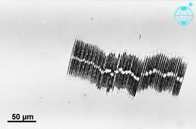
Some pennate diatoms are capable of a type of locomotion called "gliding", which allows them to move across surfaces via adhesive mucilage secreted through a seamlike structure called the raphe.[57][58] In order for a diatom cell to glide, it must have a solid substrate for the mucilage to adhere to.
Cells are solitary or united into colonies of various kinds, which may be linked by siliceous structures; mucilage pads, stalks or tubes; amorphous masses of mucilage; or by threads of chitin (polysaccharide), which are secreted through strutted processes of the cell.
_(20671468900)-cropped.jpg)

This projection of a stack of confocal images shows the diatoms' cell wall (cyan), chloroplasts (red), DNA (blue), membranes and organelles (green).
Life cycle


Reproduction and cell size
Reproduction among these organisms is asexual by
Cell division
Vegetative cells of diatoms are
A defining characteristic of all diatoms is their restrictive and bipartite silica cell wall that causes them to progressively shrink during asexual cell division. At a critically small cell size and under certain conditions,
Sexual reproduction
Most eukaryotes are capable of sexual reproduction involving meiosis. Sexual reproduction appears to be an obligatory phase in the life cycle of diatoms, particularly as cell size decreases with successive vegetative divisions[65]. Sexual reproduction involves production of gametes and the fusion of gametes to form a zygote in which maximal cell size is restored[65]. The signaling that triggers the sexual phase is favored when cells accumulate together, so that the distance between them is reduced and the contacts and/or the perception of chemical cues is facilitated[66].
An exploration of the genomes of five diatoms and one diatom transcriptome led to the identification of 42 genes potentially involved in meiosis[67]. Thus a meiotic toolkit appears to be conserved in these six diatom species[67], indicating a central role of meiosis in diatoms as in other eukaryotes.
Sperm motility
Diatoms are mostly
Degradation by microbes
Certain species of bacteria in oceans and lakes can accelerate the rate of dissolution of silica in dead and living diatoms by using
Ecology
| Part of a series on |
| Plankton |
|---|
 |


versus silicate concentration [70]
Distribution
Diatoms are a widespread group and can be found in the
Growth
Impact
The freshwater diatom Didymosphenia geminata, commonly known as Didymo, causes severe environmental degradation in water-courses where it blooms, producing large quantities of a brown jelly-like material called "brown snot" or "rock snot". This diatom is native to Europe and is an invasive species both in the antipodes and in parts of North America.[75][76] The problem is most frequently recorded from Australia and New Zealand.[77]
When conditions turn unfavourable, usually upon depletion of nutrients, diatom cells typically increase in sinking rate and exit the upper mixed layer ("bust"). This sinking is induced by either a loss of buoyancy control, the synthesis of mucilage that sticks diatoms cells together, or the production of heavy resting spores. Sinking out of the upper mixed layer removes diatoms from conditions unfavourable to growth, including grazer populations and higher temperatures (which would otherwise increase cell metabolism). Cells reaching deeper water or the shallow seafloor can then rest until conditions become more favourable again. In the open ocean, many sinking cells are lost to the deep, but refuge populations can persist near the thermocline.
Ultimately, diatom cells in these resting populations re-enter the upper mixed layer when vertical mixing entrains them. In most circumstances, this mixing also replenishes nutrients in the upper mixed layer, setting the scene for the next round of diatom blooms. In the open ocean (away from areas of continuous upwelling[78]), this cycle of bloom, bust, then return to pre-bloom conditions typically occurs over an annual cycle, with diatoms only being prevalent during the spring and early summer. In some locations, however, an autumn bloom may occur, caused by the breakdown of summer stratification and the entrainment of nutrients while light levels are still sufficient for growth. Since vertical mixing is increasing, and light levels are falling as winter approaches, these blooms are smaller and shorter-lived than their spring equivalents.
In the open ocean, the diatom (spring) bloom is typically ended by a shortage of silicon. Unlike other minerals, the requirement for silicon is unique to diatoms and it is not regenerated in the plankton ecosystem as efficiently as, for instance, nitrogen or phosphorus nutrients. This can be seen in maps of surface nutrient concentrations – as nutrients decline along gradients, silicon is usually the first to be exhausted (followed normally by nitrogen then phosphorus).
Because of this bloom-and-bust cycle, diatoms are believed to play a disproportionately important role in the export of carbon from oceanic surface waters[78][79] (see also the biological pump). Significantly, they also play a key role in the regulation of the biogeochemical cycle of silicon in the modern ocean.[72][80]
Reason for success
Diatoms are ecologically successful, and occur in virtually every environment that contains water – not only oceans, seas, lakes, and streams, but also soil and wetlands.[
Sources for collection
Diatoms can be obtained from multiple sources.
Biogeochemistry
-
 The modern oceanic silicon cycle
Fluxes are inT mol Si y−1 (28 million metric tonsof silicon per year)

Silica cycle
The diagram shows the major
Although diatoms may have existed since the
However, the precise timing of the "take-over" remains unclear, and different authors have conflicting interpretations of the fossil record. Some evidence, such as the displacement of siliceous sponges from the shelves,[88] suggests that this takeover began in the Cretaceous (146 Ma to 66 Ma), while evidence from radiolarians suggests "take-over" did not begin until the Cenozoic (66 Ma to present).[89]
-
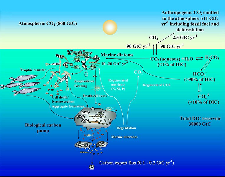 Ocean carbon cycle and diatom carbon dioxide concentration mechanisms [90]

Carbon cycle
The diagram depicts some mechanisms by which marine diatoms contribute to the
-
 Mitochondrial urea cycle in a generic diatom cell and the potential fates of urea cycle intermediates [92]

Urea cycle
A feature of diatoms is the
While often overlooked in photosynthetic organisms, the
Other
Diatoms are mainly photosynthetic; however a few are obligate
Photosynthetic diatoms that find themselves in an environment absent of oxygen and/or sunlight can switch to anaerobic respiration known as nitrate respiration (DNRA), and stay dormant for up till months and decades.[100][101]
Major
Taxonomy

.jpg)


Stephanodiscus hantzschii
.jpg)
Isthmia nervosaIsthmia nervosa

Odontella aurita
Diatoms belong to a large group of protists, many of which contain plastids rich in chlorophylls a and c. The group has been variously referred to as
Genera and species
An estimated 20,000
Classes and orders
For many years the diatoms—treated either as a class (Bacillariophyceae) or a phylum (Bacillariophyta)—were divided into just 2 orders, corresponding to the centric and the pennate diatoms (
Today (writing at mid 2020) it is recognised that the 1990 system of Round et al. is in need of revision with the advent of newer molecular work, however the best system to replace it is unclear, and current systems in widespread use such as AlgaeBase, the World Register of Marine Species and its contributing database DiatomBase, and the system for "all life" represented in Ruggiero et al., 2015, all retain the Round et al. treatment as their basis, albeit with diatoms as a whole treated as a class rather than division/phylum, and Round et al.'s classes reduced to subclasses, for better agreement with the treatment of phylogenetically adjacent groups and their containing taxa. (For references refer the individual sections below).
One proposal, by Linda Medlin and co-workers commencing in 2004, is for some of the centric diatom orders considered more closely related to the pennates to be split off as a new class, Mediophyceae, itself more closely aligned with the pennate diatoms than the remaining centrics. This hypothesis—later designated the Coscinodiscophyceae-Mediophyceae-Bacillariophyceae, or Coscinodiscophyceae+(Mediophyceae+Bacillariophyceae) (CMB) hypothesis—has been accepted by D.G. Mann among others, who uses it as the basis for the classification of diatoms as presented in Adl. et al.'s series of syntheses (2005, 2012, 2019), and also in the Bacillariophyta chapter of the 2017 Handbook of the Protists edited by Archibald et al., with some modifications reflecting the apparent non-monophyly of Medlin et al. original "Coscinodiscophyceae". Meanwhile, a group led by E.C. Theriot favours a different hypothesis of phylogeny, which has been termed the structural gradation hypothesis (SGH) and does not recognise the Mediophyceae as a monophyletic group, while another analysis, that of Parks et al., 2018, finds that the radial centric diatoms (Medlin et al.'s Coscinodiscophyceae) are not monophyletic, but supports the monophyly of Mediophyceae minus Attheya, which is an anomalous genus. Discussion of the relative merits of these conflicting schemes continues by the various parties involved.[109][110][111][112]
Adl et al., 2019 treatment
In 2019, Adl et al.[113] presented the following classification of diatoms, while noting: "This revision reflects numerous advances in the phylogeny of the diatoms over the last decade. Due to our poor taxon sampling outside of the Mediophyceae and pennate diatoms, and the known and anticipated diversity of all diatoms, many clades appear at a high classification level (and the higher level classification is rather flat)." This classification treats diatoms as a phylum (Diatomeae/Bacillariophyta), accepts the class Mediophyceae of Medlin and co-workers, introduces new subphyla and classes for a number of otherwise isolated genera, and re-ranks a number of previously established taxa as subclasses, but does not list orders or families. Inferred ranks have been added for clarity (Adl. et al. do not use ranks, but the intended ones in this portion of the classification are apparent from the choice of endings used, within the system of botanical nomenclature employed).
- Clade DiatomistaDerelle et al. 2016, emend. Cavalier-Smith 2017 (diatoms plus a subset of other ochrophyte groups)
- Phylum Diatomeae Dumortier 1821 [= BacillariophytaHaeckel 1878] (diatoms)
- Subphylum Leptocylindrophytina D.G. Mann in Adl et al. 2019
- Class Leptocylindrophyceae D.G. Mann in Adl et al. 2019 (Leptocylindrus, Tenuicylindrus)
- Class Corethrophyceae D.G. Mann in Adl et al. 2019 (Corethron)
- Subphylum Ellerbeckiophytina D.G. Mann in Adl et al. 2019 (Ellerbeckia)
- Subphylum Probosciophytina D.G. Mann in Adl et al. 2019 (Proboscia)
- Subphylum Melosirophytina D.G. Mann in Adl et al. 2019 (Aulacoseira, Melosira, Hyalodiscus, Stephanopyxis, Paralia, Endictya)
- Subphylum Coscinodiscophytina Medlin & Kaczmarska 2004, emend. (Actinoptychus, Coscinodiscus, Actinocyclus, Asteromphalus, Aulacodiscus, Stellarima)
- Subphylum Rhizosoleniophytina D.G. Mann in Adl et al. 2019 (Guinardia, Rhizosolenia, Pseudosolenia)
- Subphylum Arachnoidiscophytina D.G. Mann in Adl et al. 2019 (Arachnoidiscus)
- Subphylum Bacillariophytina Medlin & Kaczmarska 2004, emend.
- Class Mediophyceae Jouse & Proshkina-Lavrenko in Medlin & Kaczmarska 2004
- Subclass Chaetocerotophycidae Round & R.M. Crawford in Round et al. 1990, emend.
- Subclass Lithodesmiophycidae Round & R.M. Crawford in Round et al. 1990, emend.
- Subclass Thalassiosirophycidae Round & R.M. Crawford in Round et al. 1990
- Subclass Cymatosirophycidae Round & R.M. Crawford in Round et al. 1990
- Subclass Odontellophycidae D.G. Mann in Adl et al. 2019
- Subclass Chrysanthemodiscophycidae D.G. Mann in Adl et al. 2019
- Class Biddulphiophyceae D.G. Mann in Adl et al. 2019
- Subclass Biddulphiophycidae Round and R.M. Crawford in Round et al. 1990, emend.
- Biddulphiophyceae incertae sedis (Attheya)
- Class BacillariophyceaeHaeckel 1878, emend.
- Bacillariophyceae incertae sedis (Striatellaceae)
- Subclass Urneidophycidae Medlin 2016
- Subclass Fragilariophycidae Round in Round, Crawford & Mann 1990, emend.
- Subclass Bacillariophycidae D.G. Mann in Round, Crawford & Mann 1990, emend.
- Class Mediophyceae Jouse & Proshkina-Lavrenko in Medlin & Kaczmarska 2004
- Subphylum Leptocylindrophytina D.G. Mann in Adl et al. 2019
- Phylum Diatomeae Dumortier 1821 [=
See taxonomy of diatoms for more details.
Gallery
- Scanning electron microscope images
-
 Diatom Surirella spiralis
 -
 Diatoms Thalassiosira sp. on a membrane filter, pore size 0.4 μm.
 -
 Diatom Paralia sulcata.
 -
 Diatom Achanthes trinodis


-
 Stand-alone cell of Bacillaria paxillifer
 -
 Colonial group of Bacillaria paxillifer


| External videos | |
|---|---|
Three diatom species were sent to the
Evolution and fossil record
Origin
Heterokont chloroplasts appear to derive from those of red algae, rather than directly from prokaryotes as occurred in plants. This suggests they had a more recent origin than many other algae. However, fossil evidence is scant, and only with the evolution of the diatoms themselves do the heterokonts make a serious impression on the fossil record.
Earliest fossils
The earliest known fossil diatoms date from the early
Relation to grasslands
The expansion of
Relation to climate
Diatom diversity over the Cenozoic has been very sensitive to global temperature, particularly to the equator-pole temperature gradient. Warmer oceans, particularly warmer polar regions, have in the past been shown to have had substantially lower diatom diversity. Future warm oceans with enhanced polar warming, as projected in global-warming scenarios,[122] could thus in theory result in a significant loss of diatom diversity, although from current knowledge it is impossible to say if this would occur rapidly or only over many tens of thousands of years.[121]
Method of investigation
The fossil record of diatoms has largely been established through the recovery of their siliceous
Diatoms preserved in lake sediments are widely used for paleoenvironmental reconstructions of Quaternary climate, especially for closed-basin lakes which experience fluctuations in water depth and salinity.
Isotope records

When diatoms die their shells (


The initial development of the halocline and
Diversification
The Cretaceous record of diatoms is limited, but recent studies reveal a progressive diversification of diatom types. The Cretaceous–Paleogene extinction event, which in the oceans dramatically affected organisms with calcareous skeletons, appears to have had relatively little impact on diatom evolution.[136]
Turnover
Although no mass extinctions of marine diatoms have been observed during the Cenozoic, times of relatively rapid evolutionary turnover in marine diatom species assemblages occurred near the Paleocene–Eocene boundary,[137] and at the Eocene–Oligocene boundary.[138] Further turnover of assemblages took place at various times between the middle Miocene and late Pliocene,[139] in response to progressive cooling of polar regions and the development of more endemic diatom assemblages.
A global trend toward more delicate diatom frustules has been noted from the Oligocene to the Quaternary.[123] This coincides with an increasingly more vigorous circulation of the ocean's surface and deep waters brought about by increasing latitudinal thermal gradients at the onset of major ice sheet expansion on Antarctica and progressive cooling through the Neogene and Quaternary towards a bipolar glaciated world. This caused diatoms to take in less silica for the formation of their frustules. Increased mixing of the oceans renews silica and other nutrients necessary for diatom growth in surface waters, especially in regions of coastal and oceanic upwelling.
Genetics

Expressed sequence tagging
In 2002, the first insights into the properties of the
Genome sequencing

In 2004, the entire
Endosymbiotic gene transfer
Diatom genomics brought much information about the extent and dynamics of the endosymbiotic
Horizontal gene transfer
In addition to EGT, horizontal gene transfer (HGT) can occur independently of an endosymbiotic event. The publication of the P. tricornutum genome reported that at least 587 P. tricornutum genes appear to be most closely related to bacterial genes, accounting for more than 5% of the P. tricornutum proteome. About half of these are also found in the T. pseudonana genome, attesting their ancient incorporation in the diatom lineage.[145]
Genetic engineering
To understand the biological mechanisms which underlie the great importance of diatoms in geochemical cycles, scientists have used the Phaeodactylum tricornutum and Thalassiosira spp. species as model organisms since the 90's.[149] Few molecular biology tools are currently available to generate mutants or transgenic lines :
Transfected plasmids can be either randomly integrated into the diatom's chromosomes or maintained as stable circular
Furthermore, these systems now allow the use of the CRISPR-Cas genome edition tool, leading to a fast production of functional knock-out mutants[152][153] and a more accurate comprehension of the diatoms' cellular processes.
Human uses
-
 Diatomaceous earth consisting of centric (radially symmetric) and pennate (bilaterally symmetric) diatoms suspended in water.
(click 3 times to fully enlarge)

Paleontology
Decomposition and decay of diatoms leads to
Industrial
Diatoms, and their shells (frustules) as
Diatomite is considered to be a natural nano material and has many uses and applications such as: production of various ceramic products, construction ceramics, refractory ceramics, special oxide ceramics, for production of humidity control materials, used as filtration material, material in the cement production industry, initial material for production of prolonged-release drug carriers, absorption material in an industrial scale, production of porous ceramics, glass industry, used as catalyst support, as a filler in plastics and paints, purification of industrial waters, pesticide holder, as well as for improving the physical and chemical characteristics of certain soils, and other uses.[155][156][157]
Diatoms are also used to help determine the origin of materials containing them, including seawater.
Nanotechnology
The deposition of silica by diatoms may also prove to be of utility to
- Supporting and regulating services provided by marine diatoms and some of their negative impacts
-
 CNN =dimethylsulphide, DMSP = dimethylsulfoniopropionate, VOCs = volatile organic compounds
dashed arrow: negative effect, solid arrow: positive effects

Forensic
The main goal of diatom analysis in
History of discovery

The first illustrations of diatoms are found in an article from 1703 in Transactions of the Royal Society showing unmistakable drawings of Tabellaria.[163] Although the publication was authored by an unnamed English gentleman, there is recent evidence that he was Charles King of Staffordshire.[163][164] It is only 80 years later that we find the first formally identified diatom, the colonial Bacillaria paxillifera, discovered and described in 1783 by Danish naturalist Otto Friedrich Müller.[163] Like many others after him, he wrongly thought that it was an animal due to its ability to move. Even Charles Darwin saw diatom remains in dust whilst in the Cape Verde Islands, although he was not sure what they were. It was only later that they were identified for him as siliceous polygastrics. The infusoria that Darwin later noted in the face paint of Fueguinos, native inhabitants of Tierra del Fuego in the southern end of South America, were later identified in the same way. During his lifetime, the siliceous polygastrics were clarified as belonging to the Diatomaceae, and Darwin struggled to understand the reasons underpinning their beauty. He exchanged opinions with the noted cryptogamist G. H. K. Thwaites on the topic. In the fourth edition of On the Origin of Species he stated that "Few objects are more beautiful than the minute siliceous cases of the diatomaceae: were these created that they might be examined and admired under the high powers of the microscope"? and reasoned that their exquisite morphologies must have functional underpinnings rather than having been created purely for humans to admire.[165]
See also
- Highly branched isoprenoid, long-chain alkenes produced by a small number of marine diatoms
Notes
References
- ^ Dangeard, P. (1933). Traite d'Algologie. Paul Lechvalier and Fils, Paris, [1] Archived 4 October 2015 at the Wayback Machine.
- ^ Dumortier, B.-C. (1822). Commentationes botanicae. Observations botaniques, dédiées à la Société d'Horticulture de Tournay (PDF). Tournay: Imprimerie de Ch. Casterman-Dieu, Rue de pont No. 10. pp. [i], [1]-116, [1, tabl., err.] Archived from the original (PDF) on 6 October 2015 – via Algaebase.
- ^ Rabenhorst, L. Flora europaea algarum aquae dulcis et submarinae (1864–1868). Sectio I. Algas diatomaceas complectens, cum figuris generum omnium xylographice impressis (1864). pp. 1–359. Lipsiae [Leipzig]: Apud Eduardum Kummerum.
- ^ Haeckel, E. (1878). Das Protistenreich Archived 10 November 2014 at the Wayback Machine.
- ^ Engler, A. & Gilg, E. (1919). Syllabus der Pflanzenfamilien: eine Übersicht über das gesamte Pflanzensystem mit besonderer Berücksichtigung der Medizinal- und Nutzpflanzen, nebst einer Übersicht über die Florenreiche und Florengebiete der Erde zum Gebrauch bei Vorlesungen und Studien über spezielle und medizinisch-pharmazeutische Botanik, 8th ed., Gebrüder Borntraeger Verlag, Berlin, 395 p.
- Perseus Project
- Perseus Project
- Perseus Project
- ISBN 0918414083.
- ^ "The Air You're Breathing? A Diatom Made That". Live Science. 11 June 2014. Archived from the original on 30 April 2018. Retrieved 30 April 2018.
- ^ "What are Diatoms?". Diatoms of North America. Archived from the original on 25 January 2020. Retrieved 28 January 2020.
- S2CID 5672525.
- ^ "King's College London – Lake Megachad". www.kcl.ac.uk. Archived from the original on 27 November 2018. Retrieved 5 May 2018.
- S2CID 128466273.
- ^ ISBN 978-0-08-053441-1. Archivedfrom the original on 3 January 2014. Retrieved 13 November 2013.
- ^ "Gas Guzzlers". Archived from the original on 22 May 2018. Retrieved 22 May 2018.
- ^ "More on Diatoms". University of California Museum of Paleontology. Archived from the original on 4 October 2012. Retrieved 20 May 2015.
- ISBN 978-0-521-31687-3.
- PMID 25049384.
- PMID 34230473.
- ^ Functional Relationship between a Dinoflagellate Host and Its Diatom Endosymbiont | Molecular Biology and Evolution | Oxford Academic
- ISBN 978-0-12-385876-4.
- ISSN 1777-5817.
- ^ The Inner Space of the Subarctic Pacific Ocean Archived 27 October 2020 at the Wayback Machine NASA Earth Expeditions, 4 September 2018.
 This article incorporates text from this source, which is in the public domain.
- .
- doi:10.7554/eLife.26066.002..
 Material was copied from this source, which is available under a Creative Commons Attribution 4.0 International License Archived 16 October 2017 at the Wayback Machine
 - ISSN 0886-6236.
- ISSN 0031-8884.
- ^ Simonsen, R., (1979). "The diatom system: ideas on phylogeny", Bacillaria, 2: 9–71.
- ^ PMID 28686696.. Modified material was copied from this source, which is available under a Creative Commons Attribution 4.0 International License
- PMID 18654305.
- PMID 19167770.
- ^ ISBN 978-0-948737-65-7. Archivedfrom the original on 1 August 2020. Retrieved 13 November 2013.
- ^ "Glass in Nature". The Corning Museum of Glass. Archived from the original on 7 March 2013. Retrieved 19 February 2013.
- ISBN 9781770054844.
- doi:10.3389/fmicb.2017.01239.. Material was copied from this source, which is available under a Creative Commons Attribution 4.0 International License Archived 16 October 2017 at the Wayback Machine
- ^ .
- ^ "Diatoms". Archived from the original on 2 February 2016. Retrieved 13 February 2016.
- ^ The Molecular Life of Diatoms
- S2CID 216033140.
- ^ S2CID 86674657.
- S2CID 10925689.[permanent dead link]
- ^ Kroger, Nils (2007). Handbook of Biomineralization: Biological Aspects and Structure Formation. Weinheim, Germany: Wiley-VCH Verlag GmbH. pp. chapter 3.
- ^ Anonymous (1702). "Two letters from a Gentleman in the Country, relating to Mr. Leeuwenhoek's Letter in Transaction, no. 283.", Philos. Trans. R. Soc. Lond. B, 23: 1494–1501.
- ^ Rogall, E. (1939). "Ueber den feinbau der kieselmembran der diatomeen", Planta: 279-291.
- ^ ISSN 2296-7745.. Modified material was copied from this source, which is available under a Creative Commons Attribution 4.0 International License
- S2CID 4266966.
- PMID 10550045.
- PMID 11106386.
- PMID 21200414.
- PMID 23626714.
- PMID 21300899.
- PMID 28738898.
- PMID 29044150.
- ISBN 9789400710849, archived from the original(PDF) on 23 July 2018, retrieved 4 October 2019
- ISSN 1939-5590.
- PMID 10470016.
- ^ Mann, David G. (February 2010). "raphid diatoms". The Tree of Life Web Project. Archived from the original on 27 September 2019. Retrieved 27 September 2019.
- ^ ISBN 978-0-520-03400-6. Archivedfrom the original on 1 August 2020. Retrieved 14 November 2013.
- ^ Lewis, W. M., Jr. (1984). "The diatom sex clock and its evolutionary significance". The American Naturalist, 123(1): 73–80
- ^ Chepurnov VA, Mann DG, Sabbe K, Vyverman W. (2004). "Experimental studies on sexual reproduction in diatoms". In Jeon KW, ed. A Survey of Cell Biology. International Review of Cytology 237: 91–154. London.
- ^ Drebes (1977) "Sexuality". In: Werner D, ed. The Biology of Diatoms, Botanical Monographs 13 250–283. Oxford: Blackwell Scientific Publications.
- S2CID 86332060.
- S2CID 80737380.
- ^ a b Mouget JL, Gastineau R, Davidovich O, Gaudin P, Davidovich NA. Light is a key factor in triggering sexual reproduction in the pennate diatom Haslea ostrearia. FEMS Microbiol Ecol. 2009 Aug;69(2):194-201. doi: 10.1111/j.1574-6941.2009.00700.x. Epub 2009 May 6. PMID: 19486155
- ^ Scalco E, Stec K, Iudicone D, Ferrante MI, Montresor M. The dynamics of sexual phase in the marine diatom Pseudo-nitzschia multistriata (Bacillariophyceae). J Phycol. 2014 Oct;50(5):817-28. doi: 10.1111/jpy.12225. Epub 2014 Sep 14. PMID: 26988637
- ^ a b Patil S, Moeys S, von Dassow P, Huysman MJ, Mapleson D, De Veylder L, Sanges R, Vyverman W, Montresor M, Ferrante MI. Identification of the meiotic toolkit in diatoms and exploration of meiosis-specific SPO11 and RAD51 homologs in the sexual species Pseudo-nitzschia multistriata and Seminavis robusta. BMC Genomics. 2015 Nov 14;16:930. doi: 10.1186/s12864-015-1983-5. Erratum in: BMC Genomics. 2019 Jul 5;20(1):544. PMID: 26572248; PMCID: PMC4647503
- .
- PMID 23560063.
- ^ .
- ^ .
- ^ S2CID 16849373.
- PMID 26929361.
- PMID 28563010.
- Virtual Museum of Canada
- ^ "DEP Reports Didymo Discovered in the West Branch Farmington River". Archived from the original on 16 February 2015. Retrieved 27 April 2015.
- ^ "Didymo Stakeholder Update – 31 October 2008". MAF Biosecurity New Zealand www.biosecurity.govt.nz. Archived from the original on 12 February 2013. Retrieved 1 December 2013.
- ^ S2CID 4394149.
- S2CID 85054779.
- ^ S2CID 5672525.
- S2CID 86067386.
- S2CID 206507070.
- ^ a b Chamberlain, C. J. (1901) Methods in Plant Histology, University of Chicago Press.
- ISBN 978-0-262-69160-4. Retrieved 14 November 2013.
- S2CID 128401816.
- .
- .
- .
- .
- ^ PMID 33995455.. Modified material was copied from this source, which is available under a Creative Commons Attribution 4.0 International License
- ISSN 1866-3516.
- ^ S2CID 16561857.. Modified material was copied from this source, which is available under a Creative Commons Attribution 4.0 International License
- from the original on 19 February 2022. Retrieved 20 May 2021.
- "Animal-like urea cycle in ocean's tiny diatoms enables marine phytoplankton to use carbon and nitrogen from their environment". ScienceDaily (Press release). 12 May 2011.
- ^ S2CID 8593895.
- ^ S2CID 4350884.
- PMID 25302562.
- PMID 21873112.
- ISSN 0948-3055.
- ISSN 1350-0872.
- PMID 21402908.
- S2CID 227259897.
- S2CID 30911529.
- ISBN 978-0-521-36318-1. Archived from the original on 24 May 2021. Retrieved 13 November 2013.[page needed]
- ISBN 0-948737-25-5.[page needed]
- S2CID 37684109.
- .
- ^ The World Register of Marine Species lists 1,356 diatom genus names from all habitats as at July 2020, of which 1,248 are "accepted".
- ^ Queries to the World Register of Marine Species, July 2020, return 299 "fossil only" genus names, of which 285 are "accepted".
- PMID 20224747.
- PMID 20224747.
- PMID 29040712.
- ^ Medlin, L.K.; Desdevises, Y. (2020). "Review of the phylogenetic reconstruction of the diatoms using molecular tools with an analysis of a seven gene data set using multiple outgroups and morphological data for a total evidence approach" (PDF). Phycologia. in press. Archived (PDF) from the original on 20 July 2020. Retrieved 20 July 2020.
- PMID 30257078.
- PMID 28725218.. Material was copied from this source, which is available under a Creative Commons Attribution 4.0 International License Archived 16 October 2017 at the Wayback Machine
- ^ PMID 8975694.
- S2CID 4417951.
- hdl:10013/epic.12689.
- JSTOR 1514475.
- S2CID 451773.
- S2CID 140584104.
- ^ PMID 24465441.
- ^ IPCC Core Writing Team, 2007. "Climate Change 2007: Synthesis Report". 104.
- ^ .
- ^ "What Are "Proxy" Data? | National Centers for Environmental Information (NCEI) formerly known as National Climatic Data Center (NCDC)". www.ncdc.noaa.gov. Archived from the original on 8 March 2020. Retrieved 20 October 2020.
- ^ ISSN 1814-9332.. Material was copied from this source, which is available under a Creative Commons Attribution 3.0 International License Archived 23 February 2011 at the Wayback Machine
- from the original on 19 February 2022. Retrieved 15 December 2021.
- ^ from the original on 19 February 2022. Retrieved 15 December 2021.
- ISSN 0883-8305.
- ISSN 0921-8181.
- (PDF) from the original on 19 July 2018. Retrieved 15 December 2021.
- ISSN 0277-3791.
- ISSN 0277-3791.
- (PDF) from the original on 15 July 2021. Retrieved 15 December 2021.
- ISSN 0277-3791.
- ISSN 0277-3791.
- .
- ISBN 3-87429-307-6.
- ISBN 0-691-02542-8.
- S2CID 84781882.
- PMID 12114555.
- PMID 15608213.
- PMID 15665249.
- PMID 19029140.
- ^ S2CID 8593895.
- ^ S2CID 4415177.
- PMID 17350938.
- PMID 20028555.
- S2CID 11408339. Archived from the original(PDF) on 21 April 2014. Retrieved 13 January 2019.
- ^ from the original on 19 February 2022. Retrieved 20 May 2021.
- ^ S2CID 22267097.
- ^ PMID 25897682.
- ^ PMID 29298053.
- PMID 27108533.
- PMID 28725218.
- from the original on 19 February 2022. Retrieved 8 April 2020.
- ISSN 1857-8586. Archived from the originalon 18 April 2020. Retrieved 8 April 2020.
- .
- PMID 15486572.

- PMID 12902165.
- ^ Johnson, R.C. (9 April 2009). "Diatoms could triple solar cell efficiency". EE Times. Archived from the original on 31 July 2012. Retrieved 13 April 2009.
- .
- S2CID 38370984.
- ^ hdl:11336/143676.
- S2CID 201207778.
- ^ Darwin, Richard (1866). On the Origin of Species by Means of Natural Selection: Or the Preservation of Favoured Races in the Struggle for Life.
External links
- Diatom EST database, École Normale Supérieure
- Plankton*Net, taxonomic database including images of diatom species
- Life History and Ecology of Diatoms, University of California Museum of Paleontology
- Diatoms: 'Nature's Marbles', Eureka site, University of Bergen
- Diatom life history and ecology, Microfossil Image Recovery and Circulation for Learning and Education (MIRACLE), University College London
- Diatom page Archived 8 October 2009 at the Wayback Machine, Royal Botanic Garden Edinburgh
- Geometry and Pattern in Nature 3: The holes in radiolarian and diatom tests
- Diatom QuickFacts, Monterey Bay Aquarium Research Institute
- Algae image database Academy of Natural Sciences of Philadelphia(ANSP)
- Diatom taxa Academy of Natural Sciences of Philadelphia(ANSP)
- An Introduction to the Microscopical Study of Diatoms Archived 5 August 2022 at the Wayback Machine by Robert B. McLaughlin