
Dinoflagellate
| Dinoflagellate Temporal range:
| |
|---|---|

| |
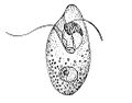
| Ceratium sp. | |
| Scientific classification | |
| Domain: | Eukaryota |
| Clade: | Diaphoretickes |
| Clade: | SAR |
| Clade: | Alveolata |
| Phylum: | Myzozoa |
| Subphylum: | Dinozoa |
| Superclass: | Dinoflagellata Bütschli 1885 [1880–1889] sensu Gomez 2012[2][3][4] |
| Classes | |
| |
| Synonyms | |
| |
The dinoflagellates (
In terms of number of species, dinoflagellates are one of the largest groups of marine eukaryotes, although substantially smaller than
About 1,555 species of free-living marine dinoflagellates are currently described.[11] Another estimate suggests about 2,000 living species, of which more than 1,700 are marine (free-living, as well as benthic) and about 220 are from fresh water.[12] The latest estimates suggest a total of 2,294 living dinoflagellate species, which includes marine, freshwater, and parasitic dinoflagellates.[2]
A rapid accumulation of certain dinoflagellates can result in a visible coloration of the water, colloquially known as
Etymology
The term "dinoflagellate" is a combination of the Greek dinos and the Latin flagellum. Dinos means "whirling" and signifies the distinctive way in which dinoflagellates were observed to swim. Flagellum means "whip" and this refers to their
History
In 1753, the first modern dinoflagellates were described by Henry Baker as "Animalcules which cause the Sparkling Light in Sea Water",[14] and named by Otto Friedrich Müller in 1773.[15] The term derives from the Greek word δῖνος (dînos), meaning whirling, and Latin flagellum, a diminutive term for a whip or scourge.
In the 1830s, the German microscopist Christian Gottfried Ehrenberg examined many water and plankton samples and proposed several dinoflagellate genera that are still used today including Peridinium, Prorocentrum, and Dinophysis.[16]
These same dinoflagellates were first defined by Otto Bütschli in 1885 as the flagellate order Dinoflagellida.[17] Botanists treated them as a division of algae, named Pyrrophyta or Pyrrhophyta ("fire algae"; Greek pyrr(h)os, fire) after the bioluminescent forms, or Dinophyta. At various times, the cryptomonads, ebriids, and ellobiopsids have been included here, but only the last are now considered close relatives. Dinoflagellates have a known ability to transform from noncyst to cyst-forming strategies, which makes recreating their evolutionary history extremely difficult.
Morphology


Dinoflagellates are unicellular and possess two dissimilar flagella arising from the ventral cell side (dinokont flagellation). They have a ribbon-like transverse flagellum with multiple waves that beats to the cell's left, and a more conventional one, the longitudinal flagellum, that beats posteriorly.
Dinoflagellates have a complex cell covering called an amphiesma or cortex, composed of a series of membranes, flattened
A transverse groove, the so-called cingulum (or cigulum) runs around the cell, thus dividing it into an anterior (episoma) and posterior (hyposoma). If and only if a theca is present, the parts are called epitheca and hypotheca, respectively. Posteriorly, starting from the transverse groove, there is a longitudinal furrow called the sulcus. The transverse flagellum strikes in the cingulum, the longitudinal flagellum in the sulcus.[25][24]
Together with various other structural and genetic details, this organization indicates a close relationship between the dinoflagellates, the Apicomplexa, and ciliates, collectively referred to as the alveolates.[23]
Dinoflagellate tabulations can be grouped into six "tabulation types":
The
Some athecate species have an internal skeleton consisting of two star-like
Theca structure and formation
The formation of thecal plates has been studied in detail through ultrastructural studies.[22]
The dinoflagellate nucleus: dinokaryon
'Core dinoflagellates' (
Classification
Generality
Dinoflagellates are protists and have been classified using both the
The peridinin dinoflagellates, named after their peridinin plastids, appear to be ancestral for the dinoflagellate lineage. Almost half of all known species have chloroplasts, which are either the original peridinin plastids or new plastids acquired from other lineages of unicellular algae through endosymbiosis. The remaining species have lost their photosynthetic abilities and have adapted to a heterotrophic, parasitic or kleptoplastic lifestyle.[33][34]
Most (but not all) dinoflagellates have a dinokaryon, described below (see: Life cycle, below). Dinoflagellates with a dinokaryon are classified under Dinokaryota, while dinoflagellates without a dinokaryon are classified under Syndiniales.
Although classified as

Jakob Schiller (1931–1937) provided a description of all the species, both marine and freshwater, known at that time.[37] Later, Alain Sournia (1973, 1978, 1982, 1990, 1993) listed the new taxonomic entries published after Schiller (1931–1937).[38][39][40][41][42] Sournia (1986) gave descriptions and illustrations of the marine genera of dinoflagellates, excluding information at the species level.[43] The latest index is written by Gómez.[2]
Identification
English-language taxonomic monographs covering large numbers of species are published for the Gulf of Mexico,[44] the Indian Ocean,[45] the British Isles,[46] the Mediterranean[47] and the North Sea.[48]
The main source for identification of freshwater dinoflagellates is the Süsswasser Flora.[49]
Calcofluor-white can be used to stain thecal plates in armoured dinoflagellates.[50]
Ecology and physiology
Habitats
Dinoflagellates are found in all aquatic environments: marine, brackish, and fresh water, including in snow or ice. They are also common in benthic environments and sea ice.
Endosymbionts
All
Nutritional strategies
Three nutritional strategies are seen in dinoflagellates:
Food inclusions contain bacteria, bluegreen algae, diatoms, ciliates, and other dinoflagellates.[54][55][56][57][58][59][60]
Mechanisms of capture and ingestion in dinoflagellates are quite diverse. Several dinoflagellates, both thecate (e.g. Ceratium hirundinella,[59] Peridinium globulus[57]) and nonthecate (e.g. Oxyrrhis marina,[55] Gymnodinium sp.[61] and Kofoidinium spp.[62]), draw prey to the sulcal region of the cell (either via water currents set up by the flagella or via pseudopodial extensions) and ingest the prey through the sulcus. In several Protoperidinium spp., e.g. P. conicum, a large feeding veil—a pseudopod called the pallium—is extruded to capture prey which is subsequently digested extracellularly (= pallium-feeding).[63][64] Oblea, Zygabikodinium, and Diplopsalis are the only other dinoflagellate genera known to use this particular feeding mechanism.[64][65][66] Katodinium (Gymnodinium) fungiforme, commonly found as a contaminant in algal or ciliate cultures, feeds by attaching to its prey and ingesting prey cytoplasm through an extensible peduncle.[67] Two related species, polykrikos kofoidii and neatodinium, shoots out a harpoon-like organelle to capture prey.[68]
Some mixotrophic dinoflagellates are able to produce neurotoxins that have anti-grazing effects on larger copepods and enhance the ability of the dinoflagellate to prey upon larger copepods. Toxic strains of K. veneficum produce karlotoxin that kills predators who ingest them, thus reducing predatory populations and allowing blooms of both toxic and non-toxic strains of K. veneficum. Further, the production of karlotoxin enhances the predatory ability of K. veneficum by immobilizing its larger prey.[69] K. arminger are more inclined to prey upon copepods by releasing a potent neurotoxin that immobilizes its prey upon contact. When K. arminger are present in large enough, they are able to cull whole populations of its copepods prey.[70]
The feeding mechanisms of the oceanic dinoflagellates remain unknown, although pseudopodial extensions were observed in Podolampas bipes.[71]
Blooms
Introduction
Dinoflagellate blooms are generally unpredictable, short, with low species diversity, and with little species succession.[72] The low species diversity can be due to multiple factors. One way a lack of diversity may occur in a bloom is through a reduction in predation and a decreased competition. The first may be achieved by having predators reject the dinoflagellate, by, for example, decreasing the amount of food it can eat. This additionally helps prevent a future increase in predation pressure by cause predators that reject it to lack the energy to breed. A species can then inhibit the growth of its competitors, thus achieving dominance.[73]
Harmful algal blooms
Dinoflagellates sometimes bloom in concentrations of more than a million cells per millilitre. Under such circumstances, they can produce toxins (generally called
_by_Noctiluca_in_Nagasaki.jpg)
A red tide occurs because dinoflagellates are able to reproduce rapidly and copiously as a result of the abundant nutrients in the water. Although the resulting red waves are an interesting visual phenomenon, they contain
Human inputs of phosphate further encourage these red tides, so strong interest exists in learning more about dinoflagellates, from both medical and economic perspectives. Dinoflagellates are known to be particularly capable of scavenging dissolved organic phosphorus for P-nutrient, several HAS species have been found to be highly versatile and mechanistically diversified in utilizing different types of DOPs.[75][76][77] The ecology of harmful algal blooms is extensively studied.[78]
Bioluminescence

At night, water can have an appearance of sparkling light due to the bioluminescence of dinoflagellates.
Dinoflagellate bioluminescence is controlled by a circadian clock and only occurs at night.[84] Luminescent and nonluminescent strains can occur in the same species. The number of scintillons is higher during night than during day, and breaks down during the end of the night, at the time of maximal bioluminescence.[85]
The luciferin-luciferase reaction responsible for the bioluminescence is pH sensitive.[83] When the pH drops, luciferase changes its shape, allowing luciferin, more specifically tetrapyrrole, to bind.[83] Dinoflagellates can use bioluminescence as a defense mechanism. They can startle their predators by their flashing light or they can ward off potential predators by an indirect effect such as the "burglar alarm". The bioluminescence attracts attention to the dinoflagellate and its attacker, making the predator more vulnerable to predation from higher trophic levels.[83]
Bioluminescent dinoflagellate ecosystem bays are among the rarest and most fragile,[86] with the most famous ones being the Bioluminescent Bay in La Parguera, Lajas, Puerto Rico; Mosquito Bay in Vieques, Puerto Rico; and Las Cabezas de San Juan Reserva Natural Fajardo, Puerto Rico. Also, a bioluminescent lagoon is near Montego Bay, Jamaica, and bioluminescent harbors surround Castine, Maine.[87] Within the United States, Central Florida is home to the Indian River Lagoon which is abundant with dinoflagellates in the summer and bioluminescent ctenophore in the winter.[88]
Lipid and sterol production
Dinoflagellates produce characteristic lipids and sterols.[89] One of these sterols is typical of dinoflagellates and is called dinosterol.
Transport
Dinoflagellate theca can sink rapidly to the seafloor in marine snow.[90]
Life cycle
Introduction
Dinoflagellates have a


Dinoflagellate cysts
The life cycle of many dinoflagellates includes at least one nonflagellated benthic stage as a

More than 10% of the approximately 2000 known marine dinoflagellate species produce cysts as part of their life cycle (see diagram on the right). These benthic phases play an important role in the ecology of the species, as part of a planktonic-benthic link in which the cysts remain in the sediment layer during conditions unfavorable for vegetative growth and, from there, reinoculate the water column when favorable conditions are restored.[94]
Indeed, during dinoflagellate evolution the need to adapt to fluctuating environments and/or to seasonality is thought to have driven the development of this life cycle stage. Most protists form dormant cysts in order to withstand starvation and UV damage.[95] However, there are enormous differences in the main phenotypic, physiological and resistance properties of each dinoflagellate species cysts. Unlike in higher plants most of this variability, for example in dormancy periods, has not been proven yet to be attributed to latitude adaptation or to depend on other life cycle traits.[96][97] Thus, despite recent advances in the understanding of the life histories of many dinoflagellate species, including the role of cyst stages, many gaps remain in knowledge about their origin and functionality.[94]
Recognition of the capacity of dinoflagellates to encyst dates back to the early 20th century, in
However, in the general life cycle of cyst-producing dinoflagellates as outlined in the 1960s and 1970s, resting cysts were assumed to be the fate of sexuality,
Yet, with the discovery that planozygotes were also able to divide it became apparent that the complexity of dinoflagellate life cycles was greater than originally thought.[103][104] Following corroboration of this behavior in several species, the capacity of dinoflagellate sexual phases to restore the vegetative phase, bypassing cyst formation, became well accepted.[105][106] Further, in 2006 Kremp and Parrow showed the dormant resting cysts of the Baltic cold water dinoflagellates Scrippsiella hangoei and Gymnodinium sp. were formed by the direct encystment of haploid vegetative cells, i.e., asexually.[107] In addition, for the zygotic cysts of Pfiesteria piscicida dormancy was not essential.[108][94]
Genomics
One of the most striking features of dinoflagellates is the large amount of cellular DNA that they contain. Most eukaryotic algae contain on average about 0.54 pg DNA/cell, whereas estimates of dinoflagellate DNA content range from 3–250 pg/cell,[31] corresponding to roughly 3000–215 000 Mb (in comparison, the haploid human genome is 3180 Mb and hexaploid Triticum wheat is 16 000 Mb). Polyploidy or polyteny may account for this large cellular DNA content,[109] but earlier studies of DNA reassociation kinetics and recent genome analyses do not support this hypothesis.[110] Rather, this has been attributed, hypothetically, to the rampant retroposition found in dinoflagellate genomes.[111][112]
In addition to their disproportionately large genomes, dinoflagellate nuclei are unique in their morphology, regulation, and composition. Their DNA is so tightly packed that exactly how many chromosomes they have is still uncertain.[113]
The dinoflagellates share an unusual mitochondrial genome organisation with their relatives, the
In most of the species, the plastid genome consist of just 14 genes.[119]
The DNA of the plastid in the peridinin-containing dinoflagellates is contained in a series of small circles called minicircles.[120] Each circle contains one or two polypeptide genes. The genes for these polypeptides are chloroplast-specific because their homologs from other photosynthetic eukaryotes are exclusively encoded in the chloroplast genome. Within each circle is a distinguishable 'core' region. Genes are always in the same orientation with respect to this core region.
In terms of DNA barcoding, ITS sequences can be used to identify species,[121] where a genetic distance of p≥0.04 can be used to delimit species,[122] which has been successfully applied to resolve long-standing taxonomic confusion as in the case of resolving the Alexandrium tamarense complex into five species.[123] A recent study[124] revealed a substantial proportion of dinoflagellate genes encode for unknown functions, and that these genes could be conserved and lineage-specific.
Evolutionary history
Dinoflagellates are mainly represented as fossils by
Molecular phylogenetics show that dinoflagellates are grouped with
The earliest stages of dinoflagellate evolution appear to be dominated by parasitic lineages, such as perkinsids and syndinians (e.g. Amoebophrya and Hematodinium).[135][136][137][138]
All dinoflagellates contain red algal plastids or remnant (nonphotosynthetic) organelles of red algal origin.[139] The parasitic dinoflagellate Hematodinium however lacks a plastid entirely.[140] Some groups that have lost the photosynthetic properties of their original red algae plastids has obtained new photosynthetic plastids (chloroplasts) through so-called serial endosymbiosis, both secondary and tertiary. Like their original plastids, the new chloroplasts in these groups can be traced back to red algae, except from those in the members of the genus Lepidodinium, which possess plastids derived from green algae, possibly Trebouxiophyceae or Ulvophyceae.[141][142] Lineages with tertiary endosymbiosis are Dinophysis, with plastids from a cryptomonad,[143] the Karenia, Karlodinium, and Takayama, which possess plastids of haptophyte origin, and the Kryptoperidiniaceae, Durinskia and Kryptoperidinium, which have plastids derived from diatoms[144][145] Some species also perform kleptoplasty.[146]
Dinoflagellate evolution has been summarized into five principal organizational types: prorocentroid, dinophysoid, gonyaulacoid, peridinioid, and gymnodinoid.[147] The transitions of marine species into fresh water have been frequent events during the diversification of dinoflagellates and have occurred recently.[148]
Many dinoflagellates also have a symbiotic relationship with cyanobacteria, called cyanobionts, which have a reduced genome and has not been found outside their hosts. The Dinophysoid dinoflagellates have two genera, Amphisolenia and Triposolenia, that contain intracellular cyanobionts, and four genera; Citharistes, Histioneis, Parahistioneis, and Ornithocercus, that contain extracellular cyanobionts.[149] Most of the cyanobionts are used for nitrogen fixation, not for photosynthesis, but some don't have the ability to fix nitrogen. The dinoflagellate Ornithocercus magnificus is host for symbionts which resides in an extracellular chamber. While it is not fully known how the dinoflagellate benefit from it, it has been suggested it is farming the cyanobacteria in specialized chambers and regularly digest some of them.[150]
Recently, the
Examples
- Alexandrium
- Gonyaulax
- Gymnodinium
- Lingulodinium polyedrum
-
 Oxyrrhea)
 -
-

-

-
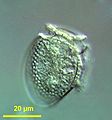
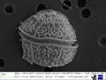 Unknown dinoflagellate under SEM (Dinophyceae)
 -

-
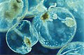
 zooxanthella, a coral endosymbiont
 -
 Noctiluca scintillans (Noctiluciphyceae)


See also
References
- PMID 21810989.
- ^ .
- PMID 25923521
- OCLC 1019558675, archivedfrom the original on 2016-05-13, retrieved 2016-09-04
- ^ OCLC 263894965.
- S2CID 83885629.
- ISBN 978-3-6421-8819-0. Archivedfrom the original on 2022-01-28. Retrieved 2020-10-22.
- S2CID 30911529.
- S2CID 86845462.
- PMID 27694774.
- ^ Gómez, F. (2005). "A list of free-living dinoflagellate species in the world's oceans". Acta Botanica Croatica. 64 (1): 129–212.
- S2CID 9810504.
- ISBN 978-0-12-385876-4.
- OCLC 722119426.
- ^ Müller, O.F. 1773. Vermium terrestrium et fluviatilium, seu Animalium Infusoriorum, Helmithicorum et Testaceorum, non marinorum, succincta historia, vol. 1. Pars prima. p. 34, 135. Faber, Havniae, et Lipsiae 1773.
- ^ Ehrenberg C.G. (1832) Beiträge zur Kenntnis der Organisation der Infusorien und ihrer geographischer Verbreitung, besonders in Sibirien. — Abhandlungen der Königlichen Akademie der Wissenschaften zu Berlin. Aus dem Jahre 1830. Physikalische Abhandlungen 1830: 1–88, Pls 1–8.
- ^ Bütschli O. (1885) 3. Unterabtheilung (Ordnung) Dinoflagellata. – In: Dr. H.G. Bronn's Klassen und Ordnungen des Thier-Reichs, wissenschaftlich dargestellt in Wort und Bild. Erster Band Protozoa. – C.F. Winter'sche Verlagshandlung, Leipzig und Heidelberg. Pp. 906–1029; Pl.
- .
- PMID 986199.
- .
- PMID 6684652.
- ^ ISBN 978-0-3231-3813-0. Archived from the original on 2014-07-07. Retrieved 2016-03-05. In Spector 1984
- ^ ISBN 978-0-1985-7747-8.
- ^ .
- S2CID 83712799.
- ISBN 978-1-86239-368-4, retrieved 2023-08-20
- PMID 21652307.
- PMID 15352316.
- ISBN 978-0-7167-1109-4.
- PMID 23159597.
- ^ ISBN 978-0-3231-3813-0. Archived from the original on 2014-07-07. Retrieved 2016-03-05. In Spector 1984
- from the original on 2018-12-14. Retrieved 2018-10-23.
- PMID 21541332.
- .
- ISBN 978-0-0805-3442-8. Archivedfrom the original on 2014-07-07. Retrieved 2016-03-05.
- ISBN 978-0-0805-3442-8. Archivedfrom the original on 2014-07-07. Retrieved 2016-03-05.
- ^ Schiller, J., 1931–1937: Dinoflagellatae (Peridinineae) in monographischer Behandlung. In: RABENHORST, L. (ed.), Kryptogamen-Flora von Deutschland, Österreichs und der Schweiz. Akad. Verlag., Leipzig. Vol. 10 (3): Teil 1 (1–3) (1931–1933): Teil 2 (1–4)(1935–1937).
- ^ Sournia A (1973). "Catalogue des espèces et taxons infraspécifiques de dinoflagellés marins actuels publiés depuis la révision de J. Schiller. I. Dinoflagellés libres". Beih. Nova Hedwigia. 48: 1–92.
- ISSN 0035-0702.
- .
- ISSN 0065-1583.
- ISSN 0181-1568.
- ^ SOURNIA, A., 1986: Atlas du Phytoplancton Marin. Vol. I: Introduction, Cyanophycées,Dictyochophycées, Dinophycées et Raphidophycées. Editions du CNRS, Paris.
- OCLC 6206528.
- OCLC 3026853.
- OCLC 681855348.
- S2CID 84744638.
- ISBN 978-3-5106-1392-2.
- ]
- S2CID 85004940.
- ^ Freudenthal et al. 2007
- OCLC 833272061.
- PMID 23194978.
- (PDF) from the original on 2022-05-15. Retrieved 2019-09-24.
- ^ S2CID 44657010.
- ISSN 0994-575X.
- ^ doi:10.1139/f61-046.
- .
- ^ S2CID 84034844.
- ^ Elbrachter M (1979). "On the taxonomy of unarmored dinophytes (Dinophyta) from the Northwest African upwelling region". Meteor Forschungsergebnisse. 30: 1–22.
- JSTOR 3225654.
- ^ Cachon PJ, Cachon M. "Le systeme stomatopharyngien de Kofoidinium Pavillard. Comparisons avec celui divers Peridiniens fibres et parasites". Protistologica. 10: 217–222.
- .
- ^ S2CID 84321400.
- .
- .
- S2CID 85988790.
- ^ "Researchers capture dinoflagellate on video shooting harpoons at prey". Archived from the original on 2019-12-28. Retrieved 2019-05-19.
- .
- PMID 22513533.
- OCLC 69377189.
- S2CID 55024118.
- .
- ISSN 0097-1618. Archived from the originalon 2007-04-30. Retrieved 2007-05-18.
- ^ S2CID 206147416.
- ^ PMID 28755722.
- ^ S2CID 3598741.
- from the original on 2014-07-07. Retrieved 2016-03-05.
- ISBN 978-0-0711-1302-1.
- PMID 8707056.
- ^ Poupin, J., A.-S. Cussatlegras, and P. Geistdoerfer. 1999. Plancton marin bioluminescent. Rapport scientifique du Laboratoire d'Océanographie de l'École Navale LOEN, Brest, France, 83 pp.
- ^ Sweeney, B. Bioluminescence and circadian rhythms. In: Taylor 1987, pp. 269–281
- ^ S2CID 3872860.
- S2CID 84990824.
- PMID 2196272.
- .
- ^ Castine Kayak (2015). "Castine Kayak Bioluminescent Bay Night Kayak Excursion". visitmaine.com. Archived from the original on 2 July 2015. Retrieved 1 July 2015.
- ^ Kennedy Duckett, Maryellen (2015-02-10). "Florida by Water: Experience Bioluminescence". National Geographic Society. Archived from the original on 2018-07-31. Retrieved 31 July 2018.
- ^ Withers, N. Dinoflagellate sterols. In: Taylor 1987, pp. 316–359
- .
- ^ from the original on 2019-02-13. Retrieved 2014-05-13.
- ISBN 978-0-6320-0915-2.
- PMID 31284474.
- ^ PMID 27694774..
 Material was copied from this source, which is available under a Creative Commons Attribution 3.0 International License Archived 2011-02-23 at the Wayback Machine
 - ^ S2CID 13213957.
- PMID 22952378.
- PMID 23717385.
- ^ Reinsch, P.F. (1905) "Die palinosphärien, ein mikroskopischer vegetabile organismus in der mukronatenkreide". ..Cent. Miner. Geol. Palaeontol..., 402–407.
- ISBN 978-0-323-13813-0.
- PMID 10978305.
- PMID 20508251.
- S2CID 85372383.
- .
- S2CID 84051080.
- S2CID 85652450.
- S2CID 85188678.
- S2CID 84228384.
- S2CID 83695348.
- ISBN 978-0-3231-3813-0. Archived from the original on 2014-07-07. Retrieved 2016-03-05. In Spector 1984
- PMID 26542574.
- PMID 28903461.
- PMID 30468510.
- ^ "Understanding relationship break-ups to protect the reef". ScienceDaily. Archived from the original on 13 May 2019. Retrieved 16 May 2019.
- PMID 22113794.
- PMID 31032404.
- PMID 12095251.
- ISBN 978-0-470-26225-2.
- PMID 22916303.
- S2CID 149458671.
- PMID 15034134.
- PMID 22916158.
- S2CID 85929661.
- PMID 28040099.
- PMID 30464192.
- ^ ISSN 0008-4026.
- PMID 9712575.
- .
- .
- ^ a b c Fensome, Robert (8 June 2022). "Dinoflagellates". AASP-The Palynological Society.[permanent dead link]
- S2CID 133043109.
- S2CID 220891202.
- hdl:2429/16056.
- .
- .
- S2CID 30051291.
- S2CID 11550698.
- S2CID 4362835.
- S2CID 28522930.
- PMID 20534454.
- PMID 25902514.
- PMID 25995366.
- hdl:10852/11559.
- .
- PMID 29066288.
- PMID 17892581..
- PMID 17227410.
- PMID 7002229.
- PMID 31369197.
- PMID 33947914.
- PMID 31235587.
- doi:10.1130/G35456.1.
- S2CID 84169394.
- PMID 23850812.
Bibliography
- Spector, D.L. (1984). Dinoflagellates. Academic Press. ISBN 978-0-3231-3813-0. Archivedfrom the original on 2014-07-07. Retrieved 2016-03-05.
- Taylor, F.J.R. (1987). The Biology of Dinoflagellates. Botanical monographs. Vol. 21. Blackwell Scientific. ISBN 978-0-6320-0915-2.
External links
- International Society for the Study of Harmful Algae
- Classic dinoflagellate monographs
- Japanese dinoflagellate site Archived 2013-05-12 at the Wayback Machine
- Noctiluca scintillans—Guide to the Marine Zooplankton of south eastern Australia, Tasmanian Aquaculture & Fisheries Institute
- Tree of Life Dinoflagellates Archived 2012-10-13 at the Wayback Machine
- Centre of Excellence for Dinophyte Taxonomy CEDiT
- Dinoflagellates Archived 2012-10-13 at the Wayback Machine
- Judson O (5 January 2010). "A Tale of Two Flagella". New York Times.
