
EcoSCOPE
This article possibly contains original research. (December 2009) |
This article is written like a personal reflection, personal essay, or argumentative essay that states a Wikipedia editor's personal feelings or presents an original argument about a topic. (December 2009) |

The ecoSCOPE is an optical sensor system, deployed from a small remotely operated vehicle (ROV) or fibre optic cable, to investigate behavior and microdistribution of small organisms in the ocean.
Deployment
Although an ROV may be very small and quiet, it is impossible to approach feeding
By imitating the long, thin snout of the garfish protruding into the security sphere of the alert herrings, an endoscope with a tip diameter of 11 mm is used. The endoscope is camouflaged to reduce the brightness-contrast against the background: the top is black and the sides are silvery. Additionally, the front of the ROV is covered by a mirror, reflecting a light gradient resembling the natural scene and making the instrument body virtually invisible to the animals. A second sensor images other copepods, phytoplankton and particles at very high magnification. Another advantage of these small "optical probes" is the minimal disruption of the current-field in the measuring volume, allowing for less disturbed surveys of microturbulence and shear.
Another video can be seen in the article for Atlantic herring.
An ecoSCOPE was also deployed to measure the dynamics of particles in a polluted estuary: see image on Particle (ecology), another as an underwater environmental monitoring system, utilizing the orientation capacity of juvenile glasseel.
-
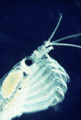
 In situ image of feeding Antarctic krill. Visible is a green spit ball and a green fecal string, important components of the biological pump.
 -
 The little tube extending from the nose is visible
 -
 Filter feeding of Antarctic krill, slowed to 1/12th of actual speed


Specifications
The ecoSCOPE is a product of the new initiative of "Ocean Online Biosensors": a synthesis of IT-sensoric and the sensing capability of ocean organisms.

Depicted in the image on the right is the central unit. On all four corners are small entrances, through which water from different sources enters (in this case, rivers and creeks in New Jersey). It flows through a small labyrinth and mixes in the central chamber. It exits through a small tube in the middle. The glasseels migrate through this small tube heading into the current. In the middle is the entrance for the eels. They test the different water qualities and migrate toward the corner, where they exit.

It is the opinion of many scientists that
The system is submerged, and a digital camera observes the exits. The dynIMAGE software monitors the frequency of decisions per exit. Many thousand of glasseels pass through the system on a single day. The three exits in the left lower corner carry water from polluted sources (one is a drinking water reservoir).
EcoSCOPE systems have already been tracking
-
 In situ image of the fibres of an Aurelia aurita from the Baltic Sea showing a prey item, probably a copepod pulled to the body by contracting the fibres in a corkscrew fashion.
 -
 Very young larvae of Atlantic herring in the typical oblique swimming position - the animal in the upper right in the classical S-shape of the beginning phase of an attack of probably a copepod - the remains of the yolk is very well visible in the transparent animal in the middle
 -
 Glasseel on the online in situ microscope at theLEOproject.
 -
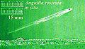
 Glasseel at the transition from ocean to freshwater; the freshwater flows from the left to the right. Glasseels are extremely difficult to image because they are transparent; see eel life history.


See also
References
- Cury PM (2004) "Tuning the ecoscope for the Ecosystem Approach to Fisheries : Perspectives on eco-system-based approaches to the management of marine resources" Marine ecology, 274: 272-275.
- Julien B, Philippe C, Pascal C and Pierre C (2008) "Safeguarding, Integrating and Disseminating Knowledge on Exploited Marine Ecosystems: The Ecoscope" International Marine Data and Information Systems, IMDIS - 2008.
- Kils, U (1992) "The ecoSCOPE and dynIMAGE: Microscale Tools for in situ Studies of Predator Prey Interaction" Archived 2008-02-21 at the Wayback Machine, Archiv für Hydrobiologie, Beihefte 36: 83-96.
- Kils, U (1994) The 3-D ecoSCOPE: A tool for investigating microdynamics and microdistributions"[usurped] In: M DeLuca (ed) Diving for Science...1994 Proceedings of the 14th Annual Scientific Diving Symposium, American Academy of Underwater Sciences. New Brunswick, New Jersey.
- Ulanowicz RE (1993) "Inventing the ecoscope", In V. Christensen and D. Pauly (eds) Trophic models of aquatic ecosystems, ICLARM Conf. Proc. 26: ix-x.
External links
- The Ecoscope Project
- Visit of President of Germany to Kiel laboratory[permanent dead link], the first propototype of the EcoSCOPE is visible in the picture, hanging from the roof.

