
Electrophysiology
Electrophysiology (from

Definition and scope
Classical electrophysiological techniques
Principle and mechanisms
Electrophysiology is the branch of physiology that pertains broadly to the flow of ions (
- Simple solid conductors, such as discs and needles (singles or arrays, often insulated except for the tip),
- Tracings on printed circuit boards or flexible polymers, also insulated except for the tip, and
- Hollow, often elongated or 'pulled', tubes filled with an electrolyte, such as glass pipettes filled with potassium chloride solution or another electrolyte solution.
The principal preparations include:
- living organisms (example in insects),
- excised tissue (acute or cultured),
- dissociated cells from excised tissue (acute or cultured),
- artificially grown cells or tissues, or
- hybrids of the above.
Neuronal electrophysiology is the study of electrical properties of biological cells and tissues within the nervous system. With neuronal electrophysiology doctors and specialists can determine how neuronal disorders happen, by looking at the individual's brain activity. Activity such as which portions of the brain light up during any situations encountered. If an electrode is small enough (micrometers) in diameter, then the
The electrophysiologist may choose not to insert the tip into a single cell. Instead, the electrode tip may be left in continuity with the extracellular space. If the tip is small enough, such a configuration may allow indirect observation and recording of
As electrode size increases, the resolving power decreases. Larger electrodes are sensitive only to the net activity of many cells, termed
Other classical electrophysiological techniques include single channel recording and amperometry.
Electrographic modalities by body part
Electrophysiological recording in general is sometimes called electrography (from
| Modality | Abbreviation | Body part | Prevalence in clinical use |
|---|---|---|---|
| electrocardiography | ECG or EKG | heart (specifically, the cardiac muscle), with cutaneous electrodes (noninvasive) | 1—very common |
| electroatriography | EAG | atrial cardiac muscle | 3—uncommon |
| electroventriculography | EVG | ventricular cardiac muscle | 3—uncommon |
| intracardiac electrogram | EGM | heart (specifically, the cardiac muscle), with intracardiac electrodes (invasive) | 2—somewhat common |
| electroencephalography | EEG | brain (usually the cerebral cortex), with extracranial electrodes | 2—somewhat common |
| electrocorticography | ECoG or iEEG | brain (specifically the cerebral cortex), with intracranial electrodes | 2—somewhat common |
| electromyography | EMG | smooth ) |
1—very common |
| electrooculography | EOG | eye—entire globe | 2—somewhat common |
| electroretinography | ERG | eye—retina specifically | 2—somewhat common |
| electronystagmography | ENG | eye—via the corneoretinal potential | 2—somewhat common |
| electroolfactography | EOG | olfactory epithelium in mammals | 3—uncommon |
| electroantennography | EAG | olfactory receptors in arthropod antennae | 4—not applicable clinically |
| electrocochleography | ECOG or ECochG | cochlea | 2—somewhat common |
| electrogastrography | EGG | stomach smooth muscle | 2—somewhat common |
electrogastroenterography |
EGEG | stomach and bowel smooth muscle | 2—somewhat common |
| electroglottography | EGG | glottis | 3—uncommon |
| electropalatography | EPG | palatal contact of tongue | 3—uncommon |
| electroarteriography | EAG | arterial flow via streaming potential detected through skin[2] | 3—uncommon |
| electroblepharography | EBG | eyelid muscle | 3—uncommon |
| electrodermography | EDG | skin | 3—uncommon |
| electropancreatography | EPG | pancreas | 3—uncommon |
| electrohysterography | EHG | uterus | 3—uncommon |
| electroneuronography | ENeG or ENoG | nerves | 3—uncommon |
| electropneumography | EPG | lungs (chest movements) | 3—uncommon |
| electrospinography | ESG | spinal cord | 3—uncommon |
| electrovomerography | EVG | vomeronasal organ | 3—uncommon |
Optical electrophysiological techniques
Optical electrophysiological techniques were created by scientists and engineers to overcome one of the main limitations of classical techniques. Classical techniques allow observation of electrical activity at approximately a single point within a volume of tissue. Classical techniques singularize a distributed phenomenon. Interest in the spatial distribution of bioelectric activity prompted development of molecules capable of emitting light in response to their electrical or chemical environment. Examples are
Intracellular recording
Intracellular recording involves measuring voltage and/or current across the membrane of a cell. To make an intracellular recording, the tip of a fine (sharp) microelectrode must be inserted inside the cell, so that the membrane potential can be measured. Typically, the resting membrane potential of a healthy cell will be -60 to -80 mV, and during an action potential the membrane potential might reach +40 mV. In 1963,
Voltage clamp

The voltage clamp technique allows an experimenter to "clamp" the cell potential at a chosen value. This makes it possible to measure how much ionic current crosses a cell's membrane at any given voltage. This is important because many of the ion channels in the membrane of a neuron are voltage-gated ion channels, which open only when the membrane voltage is within a certain range. Voltage clamp measurements of current are made possible by the near-simultaneous digital subtraction of transient capacitive currents that pass as the recording electrode and cell membrane are charged to alter the cell's potential.
Current clamp
The current clamp technique records the
Most current-clamp amplifiers provide little or no amplification of the voltage changes recorded from the cell. The "amplifier" is actually an
Patch-clamp recording

This technique was developed by Erwin Neher and Bert Sakmann who received the Nobel Prize in 1991.[5] Conventional intracellular recording involves impaling a cell with a fine electrode; patch-clamp recording takes a different approach. A patch-clamp microelectrode is a micropipette with a relatively large tip diameter. The microelectrode is placed next to a cell, and gentle suction is applied through the microelectrode to draw a piece of the cell membrane (the 'patch') into the microelectrode tip; the glass tip forms a high resistance 'seal' with the cell membrane. This configuration is the "cell-attached" mode, and it can be used for studying the activity of the ion channels that are present in the patch of membrane. If more suction is now applied, the small patch of membrane in the electrode tip can be displaced, leaving the electrode sealed to the rest of the cell. This "whole-cell" mode allows very stable intracellular recording. A disadvantage (compared to conventional intracellular recording with sharp electrodes) is that the intracellular fluid of the cell mixes with the solution inside the recording electrode, and so some important components of the intracellular fluid can be diluted. A variant of this technique, the "perforated patch" technique, tries to minimize these problems. Instead of applying suction to displace the membrane patch from the electrode tip, it is also possible to make small holes on the patch with pore-forming agents so that large molecules such as proteins can stay inside the cell and ions can pass through the holes freely. Also the patch of membrane can be pulled away from the rest of the cell. This approach enables the membrane properties of the patch to be analyzed pharmacologically. Patch-clamp may also be combined with RNA sequencing in a technique known as patch-seq by extracting the cellular contents following recording in order to characterize the electrophysiological properties relationship to gene expression and cell-type.
Sharp electrode recording
In situations where one wants to record the potential inside the cell membrane with minimal effect on the ionic constitution of the intracellular fluid a sharp electrode can be used. These micropipettes (electrodes) are again like those for patch clamp pulled from glass capillaries, but the pore is much smaller so that there is very little ion exchange between the intracellular fluid and the electrolyte in the pipette. The electrical resistance of the micropipette electrode is reduced by filling with 2-4M KCl, rather than a salt concentration which mimics the intracellular ionic concentrations as used in patch clamping.[6] Often the tip of the electrode is filled with various kinds of dyes like Lucifer yellow to fill the cells recorded from, for later confirmation of their morphology under a microscope. The dyes are injected by applying a positive or negative, DC or pulsed voltage to the electrodes depending on the polarity of the dye.
Extracellular recording
Single-unit recording
An electrode introduced into the brain of a living animal will detect electrical activity that is generated by the neurons adjacent to the electrode tip. If the electrode is a microelectrode, with a tip size of about 1 micrometre, the electrode will usually detect the activity of at most one neuron. Recording in this way is in general called "single-unit" recording. The action potentials recorded are very much like the action potentials that are recorded intracellularly, but the signals are very much smaller (typically about 1 mV). Most recordings of the activity of single neurons in anesthetized and conscious animals are made in this way. Recordings of single neurons in living animals have provided important insights into how the brain processes information. For example,
To prepare the brain for such electrode insertion, delicate slicing devices like the compresstome vibratome, leica vibratome, microtome are often employed. These instruments aid in obtaining precise, thin brain sections necessary for electrode placement, enabling neuroscientists to target specific brain regions for recording.[9]
Multi-unit recording
If the electrode tip is slightly larger, then the electrode might record the activity generated by several neurons. This type of recording is often called "multi-unit recording", and is often used in conscious animals to record changes in the activity in a discrete brain area during normal activity. Recordings from one or more such electrodes that are closely spaced can be used to identify the number of cells around it as well as which of the spikes come from which cell. This process is called spike sorting and is suitable in areas where there are identified types of cells with well defined spike characteristics. If the electrode tip is bigger still, in general the activity of individual neurons cannot be distinguished but the electrode will still be able to record a field potential generated by the activity of many cells.
Field potentials
Amperometry
(5-HT) are oxidizable. The method can also be used with cells that do not secrete oxidizable neurotransmitters by "loading" them with 5-HT or dopamine.Planar patch clamp
Planar patch clamp is a novel method developed for high throughput electrophysiology.[10] Instead of positioning a pipette on an adherent cell, cell suspension is pipetted on a chip containing a microstructured aperture. A single cell is then positioned on the hole by suction and a tight connection (Gigaseal) is formed. The planar geometry offers a variety of advantages compared to the classical experiment:
- It allows for integration of microfluidics, which enables automatic compound application for ion channel screening.
- The system is accessible for optical or scanning probe techniques.
- intracellularside can be performed.
-
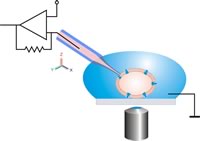 Schematic drawing of the classical patch clamp configuration. The patch pipette is moved to the cell using a micromanipulator under optical control. Relative movements between the pipette and the cell have to be avoided in order to keep the cell-pipette connection intact.
 -
 Scanning electron microscope image of a patch pipette.
 -
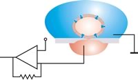 In planar patch configuration, the cell is positioned by suction. Relative movements between cell and aperture can then be excluded after sealing. An antivibration table is not necessary.
 -
 Scanning electron microscope image of a planar patch clamp chip. Both the pipette and the chip are made fromborosilicateglass.


Other methods
Solid-supported membrane (SSM)-based
With this electrophysiological approach, proteo
Bioelectric recognition assay (BERA)
The bioelectric recognition assay (BERA) is a novel method for determination of various chemical and biological molecules by measuring changes in the membrane potential of cells immobilized in a gel matrix. Apart from the increased stability of the electrode-cell interface, immobilization preserves the viability and physiological functions of the cells. BERA is used primarily in
A BERA sensor has two parts:
- The consumable biorecognition elements
- The electronic read-out device with embedded artificial intelligence.[23]
A recent advance is the development of a technique called molecular identification through membrane engineering (MIME). This technique allows for building cells with defined specificity for virtually any molecule of interest, by embedding thousands of artificial receptors into the cell membrane.[24]
Computational electrophysiology
While not strictly constituting an experimental measurement, methods have been developed to examine the conductive properties of proteins and biomembranes in silico. These are mainly molecular dynamics simulations in which a model system like a lipid bilayer is subjected to an externally applied voltage. Studies using these setups have been able to study dynamical phenomena like electroporation of membranes[25] and ion translocation by channels.[26]
The benefit of such methods is the high level of detail of the active conduction mechanism, given by the inherently high resolution and data density that atomistic simulation affords. There are significant drawbacks, given by the uncertainty of the legitimacy of the model and the computational cost of modeling systems that are large enough and over sufficient timescales to be considered reproducing the macroscopic properties of the systems themselves. While atomistic simulations may access timescales close to, or into the microsecond domain, this is still several orders of magnitude lower than even the resolution of experimental methods such as patch-clamping.[citation needed]
Clinical electrophysiology
Clinical electrophysiology is the study of how electrophysiological principles and technologies can be applied to human health. For example, clinical cardiac electrophysiology is the study of the electrical properties which govern heart rhythm and activity. Cardiac electrophysiology can be used to observe and treat disorders such as arrhythmia (irregular heartbeat). For example, a doctor may insert a catheter containing an electrode into the heart to record the heart muscle's electrical activity.
Another example of clinical electrophysiology is
Clinical reporting guidelines
See also
- Automated patch clamp
- Bioelectrochemistry
- Bioelectromagnetics
- Cardiac electrophysiology
- Clinical cardiac electrophysiology
- Clinical electrophysiology
- Clinical neurophysiology
- Electrophysiology study
- Hille equation
- History of bioelectricity
- Multiscale Electrophysiology Format
- Neurophysiology
- Slice preparation
- Transcutaneous electrical nerve stimulation
References
- S2CID 205218803.
- ^ U.S. patent 4425922A
- ^ Movie featuring Alan Hodgkin recording action potentials from a squid axon https://www.youtube.com/watch?v=k48jXzFGMc8
- PMID 30176004.
- ^ "The Nobel Prize in Physiology or Medicine 1991". nobelprize.org. Archived from the original on 10 October 2017. Retrieved 5 May 2018.
- ^ Halliwell J., Whitaker M., Ogden D. (1994) Using microelectrodes. Microelectrode techniques: the Plymouth Workshop handbook. ed. Ogden, D. Available online at http://plymsea.ac.uk/id/eprint/7954/
- PMID 14449617.
- ^ "The Nobel Prize in Physiology or Medicine 1981". nobelprize.org. Archived from the original on 23 December 2017. Retrieved 5 May 2018.
- PMID 29552595.
- ^ "Automated patch clamp" (PDF). Archived (PDF) from the original on 31 March 2010. Retrieved 17 January 2010.
- PMID 18675360.
- ^ Kintzios S., E. Pistola, P. Panagiotopoulos, M. Bomsel, N. Alexandropoulos, F. Bem, I. Biselis, R. Levin (2001) Bioelectric recognition assay (BERA). Biosensors and Bioelectronics 16: 325–36
- ^ Perdikaris, A.; Alexandropoulos, N; Kintzios, S. (2009) Development of a Novel, Ultra-rapid Biosensor for the Qualitative Detection of Hepatitis B Virus-associated Antigens and Anti-HBV, Based on "Membrane-engineered" Fibroblast Cells with Virus-Specific Antibodies and Antigens. Sensors 9: 2176–86.
- ^ Moschopoulou G.; Vitsa, K.; Bem, F.; Vassilakos, N.; Perdikaris, A.; Blouhos, P.; Yialouris, C.; Frossiniotis, D.; Anthopoulos, I.; Maggana, O.; Nomikou, K.; Rodeva, V.; Kostova, D.; Grozeva, S.; Michaelides, A.; Simonian, A.; Kintzios, S. (2008) Engineering of the membrane of fibroblast cells with virus-specific antibodies: a novel biosensor tool for virus detection. Biosensors Bioelectron. 24: 1033–36.
- ^ Flampouri E, Mavrikou S, Kintzios S, Miliaids G, Aplada-Sarli P (2010). Development and Validation of a Cellular Biosensor Detecting Pesticide Residues in Tomatoes. Talanta 80: 1799–804.
- ^ Mavrikou, S, Flampouri, E, Moschopoulou, G, Mangana, O, Michaelides, A, Kintzios, S (2008) Assessment of organophosphate and carbamate pesticide residues in cigarette tobacco with a novel cell biosensor. Sensors 8: 2818–32
- ^ Lokka K., Skandamis P., Kintzios S. (2013) Screening of Total Organophosphate Pesticides in Agricultural Products with a Cellular Biosensor CellBio 2: 131–37.
- ^ Larou, E., Yiakoumettis, I., Kaltsas, G., Petropoulos, A., Skandamis, P., Kintzios, S. (2012) High throughput cellular biosensor for the ultra-sensitive, ultra-rapid detection of aflatoxin M1. Food Control 29: 208–12
- ^ Varelas, V., Sanvicens N, Marco MP, Kintzios S (2010) Development of a cellular biosensor for the detection of 2, 4, 6- trichloroanisole (TCA). Talanta 84: 936–40
- ^ Apostolou T, Pascual N, Marco M-P, Moschos A, Petropoulos A, Kaltsas G, Kintzios S (2014) Extraction-less, rapid assay for the direct detection of 2,4,6-trichloroanisole (TCA) in cork samples. Talanta 125: 336–40.
- ^ Moschopoulou G., Kintzios S. (2006) Application of "membrane-engineering" to bioelectric recognition cell sensors for the detection of picomole concentrations of superoxide radical: a novel biosensor principle. Anal. Chimica Acta 573–74: 90–96.
- ^ Moschopoulou, G., Valero, T., Kintzios, S. (2012) Superoxide determination using membrane-engineered cells: An example of a novel concept for the construction of cell sensors with customized target recognition properties. Sens. Actuat.175: 88–94
- ^ Ferentinos K.P., C.P. Yialouris, P. Blouchos, G. Moschopoulou, V. Tsourou, Kintzios, S. (2013) Pesticide Residue Screening Using a Novel Artificial Neural Network Combined with a Bioelectric Cellular Biosensor. BioMed Research International. Article ID 813519.
- ^ Kokla A, Blouchos P., Livaniou E., Zikos C., Kakabakos S.E., Petrou P.S., Kintzios, S. (2013) Visualization of the membrane-engineering concept: evidence for the specific orientation of electroinserted antibodies and selective binding of target analytes. Journal of Molecular Recognition 26: 627–232.
- PMID 17208976.
- PMID 21843471.
- .
External links
- Book chapter on Planar Patch Clamp Archived 31 March 2010 at the Wayback Machine
Physiology types | ||
|---|---|---|
| Animals |
|  |
| Plants | ||
| Cells | ||
| Related topics |
| |