
Endochondral ossification
| Endochondral ossification | |
|---|---|
 A schematic representation of endochondral ossification. | |
| Anatomical terminology |
Endochondral ossification
Endochondral ossification is responsible for development of most bones including
Formation of the cartilage model
The initiation of endochondral ossification starts by proliferation and condensation of
| Interstitial growth | Appositional growth | |
|---|---|---|
| Cellular protagonists | Chondrocytes present within the existing cartilage. |
Chondroblasts that develop from the perichondrium.
|
| Mechanism | Chondrocytes proliferate and lay down matrix. | Chondroblasts differentiate into chondrocytes and lay down matrix. |
| Site of expansion | From within. | From the external surface of existing cartilage. |
| Outcome | Increase in length. | Increase in width and thickness. |
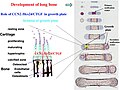
Primary center of ossification

In developing bones, ossification commences within the primary ossification center located in the center of the diaphysis (bone shaft),[5] where the following changes occur:
- The perichondrium surrounding the cartilage model transforms into the
- After the formation of the periosteum, chondrocytes in the primary center of ossification begin to grow (Matrix metalloproteinases., which causes calcification of the cartilage matrix. This calcification prevents passage of nutrients to chondrocytes leading to their death.
- Vascular endothelial growth factor (VEGF), which controls forthcoming vascular invasion.
- Alkaline phosphatase
Secondary center of ossification
During the postnatal life, a secondary ossification center appears in each end (epiphysis) of long bones. In these secondary centers, cartilage is converted to bone similarly to that occurring in a primary ossification center.[8] As the secondary ossification centers enlarge, residual cartilage persists in two distinct locations:[11]
- Articular cartilage: This layer coats the bone ends, concerned with joint movement.
- Epiphyseal growth plate: This transverse layer lies between the epiphysis and diaphysis. It’s composed of highly active chondrocytes and responsible for longitudinal bone growth. Consequently, the bone elongates at this growth plate until closure occurs at skeletal maturity.
At the end of an individual’s growth period, the production of new cartilage in the epiphyseal plate stops. After this point, existing cartilage within the plate turns into mature bone tissue.[8]
Histology

During endochondral ossification, five distinct zones can be seen at the light-microscope level:[3]
| Name | Definition |
|---|---|
| Zone of resting cartilage | This zone contains normal, resting hyaline cartilage. |
| Zone of proliferation / cell columns | In this zone, chondrocytes undergo rapid mitosis, forming distinctive looking columns. |
| Zone of maturation / hypertrophy | In this zone, the chondrocytes undergo hypertrophy (become enlarged). Chondrocytes contain large amounts of glycogen and begin to secrete vascular endothelial growth factor to initiate vascular invasion. |
| Zone of calcification | In this zone, chondrocytes are either dying or dead, leaving cavities that will later become invaded by bone-forming cells. Chondrocytes here die when they can no longer receive nutrients or eliminate wastes via diffusion. This is because the calcified matrix is much less hydrated than hyaline cartilage. |
| Zone of ossification | Osteoprogenitor cells invade the area and differentiate into osteoblasts, which elaborate matrix that becomes calcified on the surface of calcified cartilage. |

Fracture healing
For complete recovery of a fractured bone’s biomechanical functionality, the bone healing process needs to culminate in the formation of lamellar bone at the fracture site to withstand the same forces and stresses it did before the fracture. Indirect fracture healing, the most common type of bone repair,[10] relies heavily on endochondral ossification. In this type of healing, endochondral ossification occurs within the fracture gap and external to the periosteum. In contrast, intramembranous ossification takes place directly beneath the periosteum, adjacent to the broken bone’s ends.[10][12]

Additional images
-
 Masson Goldner trichrome stain of growth plate in a rabbit tibia.
 -
 Section of fetal bone of cat. ir. Irruption of the subperiosteal tissue. p. Fibrous layer of the periosteum. o. Layer of osteoblasts. im. Subperiosteal bony deposit.
 -
 Process of endochondral ossification.
 -
 Drawing of part of a longitudinal section of the developing femur of a rabbit. a. Flattened cartilage cells. b. Enlarged cartilage cells. c, d. Newly formed bone. e. Osteoblasts. f. Giant cells or osteoclasts. g, h. Shrunken cartilage cells.


References
- ^ Etymology from Greek: ἔνδον/endon, "within", and χόνδρος/chondros, "cartilage"
- ^ "Etymology of the English word endochondral". myEtymology. Archived from the original on July 14, 2011.
{{cite web}}: CS1 maint: unfit URL (link) - ^ license.
- PMID 30725884
- ^ PMID 33600952.
- ISBN 978-1975179960.
- PMID 30252246
- ^ ISBN 9781975181574.
- ^ ISBN 978-1264930395.
- ^ PMID 21489527.
- ^ PMID 37837512.
- PMID 25699016.