
Entamoeba histolytica
| Entamoeba histolytica | |
|---|---|

| |
| Entamoeba histolytica trophozoite | |
| Scientific classification | |
| Domain: | Eukaryota |
| Phylum: | Amoebozoa |
| Family: | Entamoebidae |
| Genus: | Entamoeba |
| Species: | E. histolytica
|
| Binomial name | |
| Entamoeba histolytica Schaudinn, 1903
| |

Entamoeba histolytica is an
The word histolysis literally means disintegration and dissolution of organic tissues.
Transmission
The active (trophozoite) stage exists only in the host and in fresh loose feces; cysts survive outside the host in water, in soils, and on foods, especially under moist conditions on the latter. The infection can occur when a person puts anything into their mouth that has touched the feces of a person who is infected with E. histolytica, swallows something, such as water or food, that is contaminated with E. histolytica, or swallows E. histolytica cysts (eggs) picked up from contaminated surfaces or fingers.[4] The cysts are readily killed by heat and by freezing temperatures; they survive for only a few months outside of the host.[5] When cysts are swallowed, they cause infections by excysting (releasing the trophozoite stage) in the digestive tract. The pathogenic nature of E. histolytica was first reported by Fedor A. Lösch in 1875,[1] but it was not given its Latin name until Fritz Schaudinn described it in 1903. E. histolytica, as its name suggests (histo–lytic = tissue destroying), is pathogenic; infection can be asymptomatic, or it can lead to amoebic dysentery or amoebic liver abscess.[6][7] Symptoms can include fulminating dysentery, bloody diarrhea, weight loss, fatigue, abdominal pain, and amoeboma. The amoeba can 'bore' into the intestinal wall, causing lesions and intestinal symptoms, and it may reach the blood stream or peritoneal cavity.[8] From there, it can reach vital organs of the human body, usually the liver, but sometimes the lungs, brain, and spleen.[9] A common outcome of this invasion of tissues is a liver abscess, which can be fatal if untreated.[8] Ingested red blood cells are sometimes seen in the amoeba cell cytoplasm.[10]
Risk factors
Poor sanitary conditions are known to increase the risk of contracting amebiasis E. histolytica.
Genome
The E. histolytica genome was sequenced, assembled, and automatically annotated in 2005.[17]
The genome was reassembled and reannotated in 2010.
The genome of E. histolytica has been found to have
Pathogen interaction
E. histolytica may modulate the virulence of certain human viruses and is itself a host for its own viruses.[citation needed]
For example,
A burst of research on viruses of E. histolytica stems from a series of papers published by Diamond et al. from 1972 to 1979. In 1972, they hypothesized two separate polyhedral and filamentous viral strains within E. histolytica that caused cell lysis. Perhaps the most novel observation was that two kinds of viral strains existed, and that within one type of amoeba (strain HB-301) the polyhedral strain had no detrimental effect but led to cell lysis in another (strain HK-9). Although Mattern et al. attempted to explore the possibility that these protozoal viruses could function like bacteriophages, they found no significant changes in Entamoeba histolytica virulence when infected by viruses.[25]
Immunopathogenesis
E. histolytica causes tissue destruction which leads to clinical disease. E. histolytica induces tissue damage by three main events: direct host cell death, inflammation, and parasite invasion. Once the trophozoites are excysted in the terminal ileum region, they colonize the large bowel, remaining on the surface of the mucus layer and feeding on bacteria and food particles. Occasionally, and in response to unknown stimuli, trophozoites move through the mucus layer where they come in contact with the epithelial cell layer and start the pathological process. E. histolytica has a
Diagnosis
Diagnosis is confirmed by microscopic examination for trophozoites or cysts in fresh or suitably preserved faecal specimens, smears of aspirates or scrapings obtained by proctoscopy, and aspirates of abscesses or other tissue specimen. A blood test is also available, but it is recommended only when a healthcare provider believes the infection may have spread beyond the intestine to some other organ of the body, such as the liver. However, this blood test may not be helpful in diagnosing current illness, because the test can be positive if the patient has had amebiasis in the past, even if they are not infected at the time of the test.[27] Stool antigen detection and PCR are available for diagnosis, and are more sensitive and specific than microscopy.[2]
-
 Entamoeba histolytica trophozoite
 -
 Amoebic intestinal ulcer caused by E. histolytica
 -
 erythrocytes
 -
 E. histolytica cyst
 -
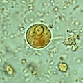 Immature E. histolytica cyst (mature cysts have 4 nuclei)
 -
 E. histolytica quadrinucleate cyst with chromatoid bodies.
 -
 Multiplication by binary fission
 -
 E. histolytica drawing
 -
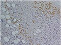 Immunohistochemical staining of trophozoites (brown) using specific anti–Entamoeba histolytica macrophage migration inhibitory factor antibodies in a patient with amebic colitis


_using_specific_anti%E2%80%93Entamoeba_histolytica_macrophage_migration_inhibitory_factor_antibodies_in_a_patient_with_amebic_colitis.jpg)
Treatment
There are a number of effective medications. Several antibiotics are available to treat Entamoeba histolytica. The infected individual will be treated with only one antibiotic if the E. histolytica infection has not made the person sick, and will most likely be prescribed two antibiotics if the person has been feeling sick.[28] Otherwise, below are other options for treatments.
Intestinal infection
Usually
Liver abscess
In addition to targeting organisms in solid tissue, primarily with drugs like metronidazole and chloroquine, treatment of liver abscess must include agents that act in the lumen of the intestine (as in the preceding paragraph) to avoid re-invasion. Surgical drainage is usually not necessary, except when rupture is imminent.[29]
People without symptoms
For people without symptoms (otherwise known as asymptomatic carriers), non-endemic areas should be treated by
| Genus and species | Entamoeba histolytica |
| Etiologic agent of: | amoebic dysentery ; extraintestinal amoebiasis, usually amoebic liver abscess; "anchovy sauce"); amoeba cutis; amoebic lung abscess ("liver-colored sputum")
|
| Infective stage | Tetranucleated cyst (having 2–4 nuclei) |
| Definitive host | Human |
| Portal of entry | Mouth |
| Mode of transmission | Ingestion of mature cyst through contaminated food or water |
| Habitat | Colon and cecum |
| Pathogenic stage | Trophozoite |
| Locomotive apparatus | Pseudopodia ("false foot”") |
| Motility | Active, progressive and directional |
| Nucleus | 'Ring and dot' appearance: peripheral chromatin and central karyosome |
| Mode of reproduction | Binary fission |
| Pathogenesis | Lytic necrosis (it looks like “flask-shaped” holes in Gastrointestinal tract sections (GIT) |
| Type of encystment | Protective and Reproductive |
| Lab diagnosis | Most common is direct fecal smear (DFS) and staining (but does not allow identification to species level); enzyme immunoassay (EIA); indirect hemagglutination (IHA); Antigen detection – monoclonal antibody; PCR for species identification. Sometimes only the use of a fixative (formalin) is effective in detecting cysts. Culture: From faecal samples – Robinson's medium, Jones' medium |
| Treatment | Diloxanide furoate (Furamide) is not commercially available in the United States or Canada (being available only from the Centers for Disease Control and Prevention). A direct comparison of efficacy showed that Paromomycin had a higher cure rate.[30] Paromomycin (Humatin) should be used with caution in patients with colitis, as it is both nephrotoxic and ototoxic. Absorption through the damaged wall of the intestinal tract can result in permanent hearing loss and kidney damage. Recommended dosage: metronidazole 750 mg three times a day orally, for 5 to 10 days followed by paromomycin 30 mg/kg/day orally in 3 equal doses for 5 to 10 days or Diloxanide furoate 500 mg 3 times a day orally for 10 days, to eradicate lumenal amoebae and prevent relapse.[31][32]
|
| Trophozoite stage | |
| Pathognomonic/diagnostic feature | Ingested RBC; distinctive nucleus |
| Cyst Stage | |
| Chromatoidal body | 'Cigar' shaped bodies (made up of crystalline ribosomes) |
| Number of nuclei | 1 in early stages, 4 when mature |
| Pathognomonic/diagnostic feature | 'Ring and dot' nucleus and chromatoid bodies |
Meiosis
In sexually reproducing eukaryotes, homologous recombination (HR) ordinarily occurs during meiosis. The meiosis-specific recombinase, Dmc1, is required for efficient meiotic HR, and Dmc1 is expressed in E. histolytica.[33] The purified Dmc1 from E. histolytica forms presynaptic filaments and catalyzes ATP-dependent homologous DNA pairing and DNA strand exchange over at least several thousand base pairs.[33] The DNA pairing and strand exchange reactions are enhanced by the eukaryotic meiosis-specific recombination accessory factor (heterodimer) Hop2-Mnd1.[33] These processes are central to meiotic recombination, suggesting that E. histolytica undergoes meiosis.[33]
Several other genes involved in both mitotic and meiotic HR are also present in E. histolytica.[34] HR is enhanced under stressful growth conditions (serum starvation) concomitant with the up-regulation of HR-related genes.[35] Also, UV irradiation induces DNA damage in E. histolytica trophozoites and activates the recombinational DNA repair pathway.[34] In particular, expression of the Rad51 protein (a recombinase) is increased about 15-fold by UV treatment.[34]
See also
- List of parasites (human)
References
- ^ S2CID 218475533.
- ^ PMID 30046644.
- PMID 9100475.
- ^ "Entamoeba histolytica". cdc.govPrevention. Center for Disease Control & Prevention. Retrieved 24 October 2017.
- ISBN 978-1-58321-403-9.
- ISBN 978-0-8385-8529-0.
- PMID 26088504.

- ^ PMID 28613582. Retrieved 11 January 2024 – via National Library of Medicine.
- PMID 33812449. Art. No. 17.
- ^ "Laboratory diagnosis of amebiasis: Entamoeba histolytica and Entamoeba dispar" (PDF). DPDx - Laboratory Identification of Parasites of Public Health Concern. Centers for Disease Control and Prevention. Retrieved 11 January 2024.
- ^ "General Information | Amebiasis | Parasites | CDC". www.cdc.gov. Retrieved 2018-03-01.
- PMID 22144440.
- PMID 28797327.
- PMID 18598643.
- PMID 20889891.
- ^ PMID 15738369.
- PMID 15729342.
- PMID 20559563.
- ISBN 978-1-904455-61-5.
- PMID 15955314.
- S2CID 30791213.
- ^ Kaur D, Gupta AK, Kumari V, Sharma R, Bhattacharya A, Bhattacharya S.
Computational prediction and validation of C/D, H/ACA and Eh_U3 snoRNAs of
Entamoeba histolytica. BMC Genomics. 2012 Aug 14;13:390. doi:
10.1186/1471-2164-13-390. PubMed PMID 22892049; PubMed Central PMCID: PMC3542256
- ^ Srivastava A, Ahamad J, Ray AK, Kaur D, Bhattacharya A, Bhattacharya S.
Analysis of U3 snoRNA and small subunit processome components in the parasitic
protist Entamoeba histolytica. Mol Biochem Parasitol. 2014 Feb;193(2):82–92. doi:
10.1016/j.molbiopara.2014.03.001. Epub 2014 Mar 12. PubMed PMID 24631428.
- PMID 2059366.
- PMID 4335522.
- PMID 30631758.
- ^ "Entamoeba histolytica". cdc.gov. Centers for Disease Control. Retrieved 24 October 2017.
- ^ "Entamoeba histolytica". Centers for Disease Control & Prevention. CDC.gov. Retrieved 24 October 2017.
- PMID 15023017.
- PMID 12397207.
- S2CID 208792864.
- ^ "Diloxanide (Systemic)". Archived from the original on 10 November 2016. Retrieved 17 November 2011.
- ^ PMID 26422142.
- ^ PMID 18402694.
- PMID 24098652.
External links
- Entamoeba histolytica image library Archived 2013-10-20 at the Wayback Machine
- Entamoeba histolytica – Centers for Disease Control and Prevention
- CDC DPDx Parasitology Diagnostic Web Site Archived 2013-07-10 at the Wayback Machine
- LSHTM 'Entamoeba Homepage
- 'Entamoeba' Genome Resource – AmoebaDB
- Entamoeba histolytica article[Bad Bug Book