Epidermal growth factor receptor
Ensembl | |||||||||
|---|---|---|---|---|---|---|---|---|---|
| UniProt | |||||||||
| RefSeq (mRNA) |
| ||||||||
| RefSeq (protein) |
| ||||||||
| Location (UCSC) | Chr 7: 55.02 – 55.21 Mb | Chr 11: 16.7 – 16.87 Mb | |||||||
| PubMed search | [3] | [4] | |||||||
| View/Edit Human | View/Edit Mouse |
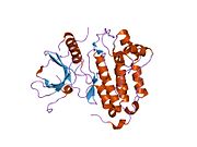
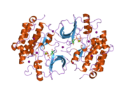
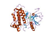
The epidermal growth factor receptor (EGFR; ErbB-1; HER1 in humans) is a
The epidermal growth factor receptor is a member of the
Epidermal growth factor and its receptor was discovered by
Deficient signaling of the EGFR and other receptor tyrosine kinases in humans is associated with diseases such as Alzheimer's, while over-expression is associated with the development of a wide variety of tumors. Interruption of EGFR signalling, either by blocking EGFR binding sites on the extracellular domain of the receptor or by inhibiting intracellular tyrosine kinase activity, can prevent the growth of EGFR-expressing tumours and improve the patient's condition[citation needed].
Function


Epidermal growth factor receptor (EGFR) is a
EGFR dimerization stimulates its intrinsic intracellular protein-tyrosine kinase activity. As a result,
Biological roles
The EGFR is essential for
Role in human disease
Cancer
Inflammatory disease
Aberrant EGFR signaling has been implicated in psoriasis, eczema and atherosclerosis.[23][24] However, its exact roles in these conditions are ill-defined.
Monogenic disease
A single child displaying multi-organ epithelial inflammation was found to have a homozygous loss of function mutation in the EGFR gene. The pathogenicity of the EGFR mutation was supported by in vitro experiments and functional analysis of a skin biopsy. His severe phenotype reflects many previous research findings into EGFR function. His clinical features included a papulopustular rash, dry skin, chronic diarrhoea, abnormalities of hair growth, breathing difficulties and electrolyte imbalances.[25]
Wound healing and fibrosis
EGFR has been shown to play a critical role in TGF-beta1 dependent fibroblast to myofibroblast differentiation.[26][27] Aberrant persistence of myofibroblasts within tissues can lead to progressive tissue fibrosis, impairing tissue or organ function (e.g. skin hypertrophic or keloid scars, liver cirrhosis, myocardial fibrosis, chronic kidney disease).
Medical applications
Drug target
The identification of EGFR as an
Many therapeutic approaches are aimed at the EGFR. Cetuximab and
Another method is using small molecules to inhibit the EGFR tyrosine kinase, which is on the cytoplasmic side of the receptor. Without kinase activity, EGFR is unable to activate itself, which is a prerequisite for binding of downstream adaptor proteins. Ostensibly by halting the signaling cascade in cells that rely on this pathway for growth, tumor proliferation and migration is diminished.
inhibitors.There are several quantitative methods available that use protein phosphorylation detection to identify EGFR family inhibitors.[36]
New drugs such as
However, many patients develop resistance. Two primary sources of resistance are the T790M mutation and
The most common adverse effect of EGFR inhibitors, found in more than 90% of patients, is a papulopustular rash that spreads across the face and torso; the rash's presence is correlated with the drug's antitumor effect.[38] In 10% to 15% of patients the effects can be serious and require treatment.[39][40]
Some tests are aiming at predicting benefit from EGFR treatment, as Veristrat.[41]
Laboratory research using genetically engineered stem cells to target EGFR in mice was reported in 2014 to show promise.[42] EGFR is a well-established target for monoclonal antibodies and specific tyrosine kinase inhibitors.[43]
Target for imaging agents
Imaging agents have been developed which identify EGFR-dependent cancers using labeled EGF.[44] The feasibility of in vivo imaging of EGFR expression has been demonstrated in several studies.[45][46]
It has been proposed that certain computed tomography findings such as ground-glass opacities, air bronchogram, spiculated margins, vascular convergence, and pleural retraction can predict the presence of EGFR mutation in patients with non-small cell lung cancer.[47]
Interactions
Epidermal growth factor receptor has been shown to
- AR,[48][49]
- ARF4,[50]
- CAV1,[51]
- CAV3,[51]
- CBLC,[58][59]
- CD44,[26]
- CDC25A,[60]
- DCN,[65][66]
- EGF,[67][68]
- GRB14,[69]
- JAK2,[77]
- NCK1,[70][80][81]
- NCK2[70][82][83]
- PKC alpha,[84]
- PLCG1,[52][85]
- PLSCR1,[86]
- PTPN1,[87][88]
- PTPN11,[57][89]
- PTPN6,[89][90]
- PTPRK,[91]
- SH2D3A,[92]
- SH3KBP1,[93][94]
- SHC1,[57][95]
- SOS1,[75][96][97]
- STAT1,[77][100]
- STAT3,[77][101]
- STAT5A,[57][77]
- UBC,[54][55][102] and
- WAS,[103]
- PAR2.[104]
In fruitflies, the epidermal growth factor receptor interacts with Spitz.[105]
References
- ^ a b c GRCh38: Ensembl release 89: ENSG00000146648 – Ensembl, May 2017
- ^ a b c GRCm38: Ensembl release 89: ENSMUSG00000020122 – Ensembl, May 2017
- ^ "Human PubMed Reference:". National Center for Biotechnology Information, U.S. National Library of Medicine.
- ^ "Mouse PubMed Reference:". National Center for Biotechnology Information, U.S. National Library of Medicine.
- PMID 15142631.
- PMID 17671639.
- ^ note, a full list of the ligands able to activate EGFR and other members of the ErbB family is given in the ErbB article)
- PMID 3494473.
- PMID 24758840.
- PMID 19959837.
- S2CID 4332354.
- PMID 16729045.
- PMID 9751121.
- S2CID 13229645.
- PMID 18470483.
- S2CID 30782707.
- PMID 8959346.
- PMID 19716155.
- ISBN 9781437717815.
- PMID 15118073.
- S2CID 11790891.
- PMID 25394504.
- PMID 11056418.
- PMID 16076471.
- PMID 24691054.
- ^ PMID 23589287.
- PMID 24134702.
- PMID 15118125.
- PMID 36267895.
- PMID 24533047.
- ^ PMID 36817181.
- S2CID 45076898.
- PMID 16235569.
- PMID 20387330.
- ^ Patel N (11 May 2015). "Cuba Has a Lung Cancer Vaccine—And America Wants It". Wired. Retrieved 13 May 2015.
- S2CID 30003827.
- ^ PMID 19671843.
- PMID 23383079.
- PMID 22472354.
- S2CID 7782594.
- S2CID 44854672.
- PMID 25346520.
- PMID 24269963.
- doi:10.1002/jrs.4678.
- .
- PMID 27748899.
- ^ Herrera Ortiz AF, Cadavid Camacho T, Vásquez Perdomo A, Castillo Herazo V, Arambula Neira J, Yepes Bustamante M, Cadavid Camacho E. Clinical and CT patterns to predict EGFR mutation in patients with non-small cell lung cancer: A systematic literature review and meta-analysis. European Journal of Radiology Open.2022;9:100400. https://doi.org/10.1016/j.ejro.2022.100400
- S2CID 46121331.
- S2CID 23831527.
- PMID 12446727.
- ^ PMID 9374534.
- ^ PMID 12061819.
- ^ PMID 10086340.
- ^ PMID 18316398.
- ^ PMID 18508924.
- PMID 18273061.
- ^ PMID 16729043.
- PMID 10571044.
- S2CID 28195948.
- PMID 11912208.
- PMID 9642287.
- PMID 9535896.
- PMID 11950845.
- S2CID 10127053.
- PMID 12105206.
- PMID 9988678.
- ^ PMID 10085134.
- PMID 12093292.
- ^ PMID 8647858.
- ^ PMID 10026169.
- S2CID 26838266.
- S2CID 23869665.
- PMID 7527043.
- S2CID 10640461.
- ^ PMID 7510700.
- PMID 1322798.
- ^ PMID 10358079.
- PMID 11278868.
- PMID 11483589.
- PMID 9362449.
- PMID 1333047.
- PMID 9737977.
- PMID 9843575.
- PMID 12878187.
- S2CID 24571987.
- PMID 12009895.
- PMID 10889023.
- PMID 8621392.
- ^ PMID 7673163.
- PMID 9733788.
- PMID 15899872.
- PMID 10187783.
- S2CID 635702.
- PMID 12177062.
- PMID 9544989.
- PMID 10675333.
- PMID 9447973.
- PMID 10777553.
- S2CID 26366427.
- PMID 12070153.
- PMID 15485908.
- PMID 18632619.
- PMID 9307968.
- S2CID 227135487.
- PMID 12648473.
Further reading
- Carpenter G (1987). "Receptors for epidermal growth factor and other polypeptide mitogens". Annual Review of Biochemistry. 56 (1): 881–914. PMID 3039909.
- Boonstra J, Rijken P, Humbel B, Cremers F, Verkleij A, van Bergen en Henegouwen P (May 1995). "The epidermal growth factor". Cell Biology International. 19 (5): 413–30. S2CID 20186286.
- Carpenter G (August 2000). "The EGF receptor: a nexus for trafficking and signaling". BioEssays. 22 (8): 697–707. S2CID 767308.
- Filardo EJ (February 2002). "Epidermal growth factor receptor (EGFR) transactivation by estrogen via the G-protein-coupled receptor, GPR30: a novel signaling pathway with potential significance for breast cancer". The Journal of Steroid Biochemistry and Molecular Biology. 80 (2): 231–8. S2CID 34995614.
- Tiganis T (January 2002). "Protein tyrosine phosphatases: dephosphorylating the epidermal growth factor receptor". IUBMB Life. 53 (1): 3–14. S2CID 8376444.
- Di Fiore PP, Scita G (October 2002). "Eps8 in the midst of GTPases". The International Journal of Biochemistry & Cell Biology. 34 (10): 1178–83. PMID 12127568.
- Benaim G, Villalobo A (August 2002). "Phosphorylation of calmodulin. Functional implications" (PDF). European Journal of Biochemistry. 269 (15): 3619–31. PMID 12153558.
- Leu TH, Maa MC (January 2003). "Functional implication of the interaction between EGF receptor and c-Src". Frontiers in Bioscience. 8 (1–3): s28–38. S2CID 20827945.
- Anderson NL, Anderson NG (November 2002). "The human plasma proteome: history, character, and diagnostic prospects". Molecular & Cellular Proteomics. 1 (11): 845–67. PMID 12488461.
- Kari C, Chan TO, Rocha de Quadros M, Rodeck U (January 2003). "Targeting the epidermal growth factor receptor in cancer: apoptosis takes center stage". Cancer Research. 63 (1): 1–5. PMID 12517767.
- Bonaccorsi L, Muratori M, Carloni V, Zecchi S, Formigli L, Forti G, Baldi E (February 2003). "Androgen receptor and prostate cancer invasion". International Journal of Andrology. 26 (1): 21–5. PMID 12534934.
- Reiter J, Maihle NJ (May 2003). "Characterization and expression of novel 60-kDa and 110-kDa EGFR isoforms in human placenta". Annals of the New York Academy of Sciences. 995 (1): 39–47. S2CID 9377682.
- Adams TE, McKern NM, Ward CW (June 2004). "Signalling by the type 1 insulin-like growth factor receptor: interplay with the epidermal growth factor receptor". Growth Factors. 22 (2): 89–95. S2CID 86844427.
- Ferguson KM (November 2004). "Active and inactive conformations of the epidermal growth factor receptor". Biochemical Society Transactions. 32 (Pt 5): 742–5. PMID 15494003.
- Chao C, Hellmich MR (December 2004). "Bi-directional signaling between gastrointestinal peptide hormone receptors and epidermal growth factor receptor". Growth Factors. 22 (4): 261–8. S2CID 35208079.
- Carlsson J, Ren ZP, Wester K, Sundberg AL, Heldin NE, Hesselager G, Persson M, Gedda L, Tolmachev V, Lundqvist H, Blomquist E, Nistér M (March 2006). "Planning for intracavitary anti-EGFR radionuclide therapy of gliomas. Literature review and data on EGFR expression". Journal of Neuro-Oncology. 77 (1): 33–45. S2CID 42293693.
- Scartozzi M, Pierantoni C, Berardi R, Antognoli S, Bearzi I, Cascinu S (April 2006). "Epidermal growth factor receptor: a promising therapeutic target for colorectal cancer". Analytical and Quantitative Cytology and Histology. 28 (2): 61–8. PMID 16637508.
- Prudkin L, Wistuba II (October 2006). "Epidermal growth factor receptor abnormalities in lung cancer. Pathogenetic and clinical implications". Annals of Diagnostic Pathology. 10 (5): 306–15. PMID 16979526.
- Ahmed SM, Salgia R (November 2006). "Epidermal growth factor receptor mutations and susceptibility to targeted therapy in lung cancer". Respirology. 11 (6): 687–92. S2CID 38429131.
- Zhang X, Chang A (March 2007). "Somatic mutations of the epidermal growth factor receptor and non-small-cell lung cancer". Journal of Medical Genetics. 44 (3): 166–72. PMID 17158592.
- Mellinghoff IK, S2CID 15077838.
- Nakamura JL (April 2007). "The epidermal growth factor receptor in malignant gliomas: pathogenesis and therapeutic implications". Expert Opinion on Therapeutic Targets. 11 (4): 463–72. S2CID 21947310.
External links
- Epidermal Growth Factor Receptor at the U.S. National Library of Medicine Medical Subject Headings (MeSH)
- Overview of all the structural information available in the PDB for UniProt: P00533 (Human Epidermal growth factor receptor) at the PDBe-KB.


