
Epidermoid cyst
| Epidermoid cyst | |
|---|---|
 | |
| Epidermal cyst on the neck, inflamed | |
| Specialty | Dermatology |

An epidermoid cyst or epidermal inclusion cyst
Signs and symptoms
The epidermoid cyst may have no symptoms, or it may be painful when touched. It can release macerated
Although they are not
Diagnosis
Epidermoid cysts are usually diagnosed when a person notices a bump on their skin and seeks medical attention. The definitive diagnosis is made after excision by a
Treatment
Cysts can be removed by excision.[6]
In case of fronto-ethmoidal epidermoid cysts, surgical resection appears to be the mainstay of treatment; however, the extent of resection is dictated by adherence of the tumor capsule to the surrounding vital structures.[7]
Hydrogen peroxide gel (H2O2) was previously recommended for cyst treatment, particularly those on body piercings. However the gel cannot adequately permeate the cyst and was not found to be effective.[8] Hydrogen peroxide is no longer recommended for wound care by doctors as it can damage the healing tissues.[9]
On body piercings, self treatment with a hot saline soak to help drain the cyst and the use of an antibacterial or medicated
Terminology
Several synonyms exist for epidermoid cysts, including epidermal cyst, infundibular cyst, keratin cyst and epidermal inclusion cyst
Epidermoid cyst may be classified as a sebaceous cyst,[15] although technically speaking it is not sebaceous.[16] "True" sebaceous cysts, cysts which originate from sebaceous glands and which contain sebum, are relatively rare and are known as steatocystoma simplex or, if multiple, as steatocystoma multiplex. Medical professionals have suggested that the term sebaceous cyst be avoided since it can be misleading.[17]: 31 In practice, however, the term is still often used for epidermoid and pilar cysts.
Additional images
-
 Epidermal inclusion cyst on the nape of a person's neck
 -
 Epidermal cyst in the earlobe


-
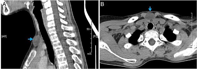 CT scan, showing a homogenous hypodense volume (unspecific cyst-like)
 -
 Epidermoid cyst in a testicle on ultrasound, with lamellated ("onion skin") appearance
 -
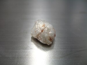 Macroscopic appearance of a resected (surgically removed) intracranial cyst, with pearl appearance
 -
 Surgery of a suprasternal epidermoid cyst, showing a smooth surface
 -
 Histopathology, showing a keratinizing stratified squamous epithelium, and a lumen containing keratin flakes
 -
 Histopathology showing epithelium and lamellated keratin (left)


See also
- Intracranial epidermoid cyst
- List of cutaneous neoplasms associated with systemic syndromes
- Proliferating epidermoid cyst
- Verrucous cyst
References
- ^ "Epidermal inclusion cyst information Diseases Database". www.diseasesdatabase.com. 17 February 2018.
- ^ "cysts - British Association of Dermatologists". Archived from the original on 2008-01-10. Retrieved 2007-11-14.
- ISBN 1-85233-912-8. Retrieved March 2, 2018.
- PMID 17847698.
- PMID 29681810.
- PMID 11996427. Archived from the originalon 2008-07-06. Retrieved 2007-11-15.
- ProQuest 922251775.
- PMID 14608258.
- PMID 11225528.
- ^ "Cysts | the Body Poetry Piercing Clinic". Archived from the original on 2014-04-23. Retrieved 2014-04-21.
- ^ Melton, Jason R. Swanson and Jeffrey L. "Epidermoid cyst". www.meddean.luc.edu.
- ISBN 0-07-138076-0.
- ISBN 0-7216-2921-0.
- ^ "Epidermoid cyst". Retrieved 2007-11-14.
- ^ "Epidermoid and pilar cysts (previously known as sebaceous cysts)". British Association of Dermatologists. Archived from the original on February 5, 2016. Retrieved April 2, 2014.
- ^ "Epidermoid and Pilar Cysts (Sebaceous Cysts) - Patient UK". Archived from the original on 2013-07-06. Retrieved 2013-03-04.
- ISBN 978-0-7216-9003-2.