
Epididymis
| Epididymis | |
|---|---|
Wolffian duct | |
| Vein | Pampiniform plexus |
| Identifiers | |
| Latin | epididymis |
| MeSH | D004822 |
| TA98 | A09.3.02.001 |
| TA2 | 3603 |
| FMA | 18255 |
| Anatomical terminology] | |
The epididymis (/ɛpɪˈdɪdɪmɪs/; pl.: epididymides /ɛpɪdɪˈdɪmədiːz/ or /ɛpɪˈdɪdəmɪdiːz/) is an elongated tubular structure attached to the posterior side of each one of the two male reproductive glands, the testicles. It is a single, narrow, tightly coiled tube in adult humans, 6 to 7 centimetres (2.4 to 2.8 in) in length; uncoiled the tube would be approximately 6 m (20 feet) long.[1] It connects the testicle to the vas deferens in the male reproductive system. The epididymis serves as an interconnection between the multiple efferent ducts at the rear of a testicle (proximally), and the vas deferens (distally). Its primary function is the storage, maturation and transport of sperm cells.
Structure
The epididymis is situated posterior and somewhat lateral to the testis. The epididymis is invested completely by the tunica vaginalis (which is continuous with the tunica vaginalis covering the testis).[2]: 1296
The epididymis can be divided into three main regions:
- The head (histologically by a thick epithelium with long stereocilia (described below) and a little smooth muscle.[3] It is involved in absorbing fluid to make the spermmore concentrated. The concentration of the sperm here is dilute.
- The body (Latin: corpus). This has an intermediate epithelium and smooth muscle thickness.[3]
- The tail (Latin: cauda). This has the thinnest epithelium of the three regions and the greatest quantity of smooth muscle.ductus deferens (s. vas deferens).[2]: 1296
Histology
The epididymis is covered by a two layered
- Principal cells: columnar cells that, with the basal cells, form the majority of the epithelium. In the caput (head) region these cells have long stereocilia that are tuft-like extensions that project into the glycerylphosphorylcholineinto the lumen.
- Basal cells: shorter, pyramid-shaped cells, which contact the basal lamina but taper off before their apical surfaces reach the lumen. These are thought to be undifferentiated precursors of principal cells.
- Apical cells: predominantly found in the head region
- Clear cells: predominant in the tail region
- Intraepithelial lymphocytes: distributed throughout the tissue.
- Intraepithelial macrophages[5][6]
Stereocilia
The stereocilia of the epididymis are long cytoplasmic projections that have an actin filament backbone.[4] These filaments have been visualized at high resolution using fluorescent phalloidin that binds to actin filaments.[4] The stereocilia in the epididymis are non-motile. These membrane extensions increase the surface area of the cell, allowing for greater absorption and secretion. It has been shown that epithelial sodium channel
Because sperm are initially non-motile as they leave the
Development
In the embryo, the epididymis develops from tissue that once formed the mesonephros, a primitive kidney found in many aquatic vertebrates. Persistence of the cranial end of the mesonephric duct will leave behind a remnant called the appendix of the epididymis. In addition, some mesonephric tubules can persist as the paradidymis, a small body caudal to the efferent ductules.
The epoophoron is a homologous remnant in the female.
Function
Role in storage of sperm and ejaculant
Spermatozoa formed in the testis enter the caput epididymis, progress to the corpus, and finally reach the cauda region, where they are stored. Sperm entering the caput epididymis are incomplete—they lack the ability to swim forward (motility) and to
The epididymis secretes immobilin, a large glycoprotein that is responsible for the creating of the viscoelastic luminal environment that serves to mechanically immobilize spermatozoa until ejaculation. Immobilin is predominantly secreted into the proximal caput epididymis prior to the acquisition of the potential for sperm motility.[9]
During emission, sperm flow from the cauda epididymis (which functions as a storage reservoir) into the vas deferens where they are propelled by the
Contrary to popular belief, sperm are capable of causing a pregnancy even without ever travelling through the epididymis.[10][11] This has been proven in two cases in the United States in the 1980s where a couple of men's vasa deferentia were directly surgically attached to their efferent ducts and these men both subsequently impregnated their partners within the next couple of years.[10] This has also been proven in a similar case in Western Europe in the early 1990s.[11]
Antioxidant defenses
During their transit through the epididymis, the
Clinical significance
Inflammation
An inflammation of the epididymis is called epididymitis. It is much more common than testicular inflammation, termed orchitis.
Surgical removal
Epididymotomy is the placing of an incision into the epididymis and is sometimes considered as a treatment option for acute suppurating epididymitis.
Epididymectomy is the surgical removal of the epididymis sometimes performed for post-vasectomy pain syndrome and for refractory cases of epididymitis.
Epididymectomy is also performed for
Other animals
The epididymis is present in all male reptiles, birds, and mammals.[1] The caput epididymis is fused to the testis in eutherian mammals, but not in marsupials.[13]
In reptiles, there is an additional canal between the testis and the head of the epididymis and which receives the various efferent ducts. This is, however, absent in all birds and mammals.[14]
Gallery
-
 Human male reproductive system
 -
 Testis
 -
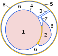 Schematic drawing: cross-section through a testicle
 -
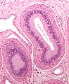 Micrograph of epididymis - H&E stain
 -
 Micrograph
 -
 Deep dissection of epididymis


![[1]](https://commons.m.wikimedia.org/wiki/File:Epididymal_head,_with_measurement,_longitudinal_view.png)
See also
- Epididymis evolution from reptiles to mammals
- Epididymal hypertension– Condition that arises during male sexual arousal when seminal fluid is not ejaculated
- List of distinct cell types in the adult human body
Notes
- ^ ISBN 978-0-7817-9141-0.
- ^ OCLC 1201341621.)
{{cite book}}: CS1 maint: location missing publisher (link) CS1 maint: others (link - ^ ISBN 978-0470958513.
- ^ S2CID 58026884.
- PMID 21310816.
- PMID 24648397.
- PMID 19136456.
- PMID 10194636.
- PMID 29541061.
- ^ PMID 3338593.
- ^ PMID 8062964.
- ^ S2CID 143424672.
- ISBN 978-1-4863-0753-1.
- ISBN 0-03-910284-X.
External links
- Histology image: 16903loa – Histology Learning System at Boston University