
Ethmoid bone
| Ethmoid bone | |
|---|---|
 Ethmoid inside the orbit (brown) | |
 Animation of the ethmoid bone | |
| Details | |
| Identifiers | |
| Latin | os ethmoidale |
| MeSH | D005004 |
| TA98 | A02.1.07.001 |
| TA2 | 721 |
| FMA | 52740 |
| Anatomical terms of bone | |
The ethmoid bone (. The cubical bone is lightweight due to a spongy construction. The ethmoid bone is one of the bones that make up the orbit of the eye.
Structure
The ethmoid bone is an anterior cranial bone located between the eyes.
Articulations
The ethmoid
- two bones of the neurocranium—the frontal, and the sphenoid (at the sphenoidal body and at the sphenoidal conchae).
- eleven bones of the viscerocranium—, two .
Development
The ethmoid is
The labyrinths are first developed, ossific granules making their appearance in the region of the
At birth, the bone consists of the two labyrinths, which are small and ill-developed. During the first year after birth, the perpendicular plate and crista galli begin to ossify from a single center, and are joined to the labyrinths about the beginning of the second year.
The cribriform plate is ossified partly from the perpendicular plate and partly from the labyrinths.
The development of the ethmoidal cells begins during fetal life.
Function
Role in magnetoception
Some
Clinical significance
Fracture of the
The porous fragile nature of the ethmoid bone makes it particularly susceptible to fractures. The ethmoid is usually fractured from an upward force to the nose. This could occur by hitting the dashboard in a car crash or landing on the ground after a fall. The ethmoid fracture can produce bone fragments that penetrate the cribriform plate. This trauma can lead to a leak of cerebrospinal fluid into the nasal cavity. These openings let opportunistic bacteria in the nasal cavity enter the sterile environment of the central nervous system (CNS). The CNS is usually protected by the blood–brain barrier, but holes in the cribriform plate let bacteria get through the barrier. The blood–brain barrier makes it extremely difficult to treat such infections, because only certain drugs can cross into the CNS.
An ethmoid fracture can also sever the olfactory nerve. This injury results in anosmia (loss of smell). A reduction in the ability to taste is also a side effect because it is based so heavily on smell. This injury is not fatal, but can be dangerous, as when a person fails to smell smoke, gas, or spoiled food.[3] In fact, people with anosmia were more than four times as likely to die in five years compared to those with a healthy sense of smell.[8]
This section needs expansion. You can help by adding to it. (December 2013) |
Additional images
-
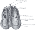 Ethmoid bone from above.
 -
 Perpendicular plate of ethmoid.
 -
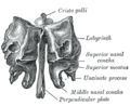 Ethmoid bone (view from behind).
 -
 Ethmoid bone from the right side.
 -
 Side view of the skull.


See also
References
![]() This article incorporates text in the public domain from page 153 of the 20th edition of Gray's Anatomy (1918)
This article incorporates text in the public domain from page 153 of the 20th edition of Gray's Anatomy (1918)
- OED2nd edition, 1989 as /ˈεθmɔɪd/.
- ^ Entry "ethmoid" in Merriam-Webster Online Dictionary.
- ^ ISBN 978-0-07-340371-7.
- ^ "Ethmoid bone". www.anatomynext.com. Archived from the original on 3 September 2020. Retrieved 1 March 2018.
- ^ Fehrenbach; Herring (2012). Illustrated Anatomy of the Head and Neck. Elsevier. p. 52.
- ^ Jacobs (2008). Human Anatomy. Elsevier. p. 210.
- S2CID 2385818.
- PMID 25271633.
Further reading
- Saladin, Kenneth S. (2010). Anatomy and Physiology: the Unity of Form and Function (5th ed.). New York: McGraw Hill. ISBN 978-0-07-128341-0.
- Banks, Peter; Brown, Andrew E. (2000). Fractures of the facial skeleton. Oxford: Wright. ISBN 0-7236-1034-7.
External links
- http://www.theregister.com/2006/11/17/the_odd_body_nose_compass/
- "Anatomy diagram: 34256.000-1". Roche Lexicon – illustrated navigator. Elsevier. Archived from the original on 27 December 2012.
| National | |
|---|---|
| Other | |