
Fragment antigen-binding

The fragment antigen-binding region (Fab region) is a region on an antibody that binds to antigens. It is composed of one constant and one variable domain of each of the heavy and the light chain. The variable domain contains the paratope (the antigen-binding site), comprising a set of complementarity-determining regions, at the amino terminal end of the monomer. Each arm of the Y thus binds an epitope on the antigen.
Preparation
In an experimental setting,
-
 Heavy and light chains, variable and constant regions of an antibody.
 -
 An antibody digested by papain yields three fragments: two Fab fragments and one Fc fragment
 -
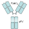 An antibody digested by pepsin yields two fragments: a F(ab')2 fragment and a pFc' fragment


The variable regions of the heavy and light chains can be fused together to form a single-chain variable fragment (scFv), which is only half the size of the Fab fragment, yet retains the original specificity of the parent immunoglobulin.[2]
Applications
Theraputics
Fabs have seen some therapeutic use in
Fabs are a common form-factor for
Diagnostics
Fab antibodies also have diagnostic use.
Fab fragments are often fused to small proteins (<100 kDa) that have lower scattering, resulting in images with less contrast.
See also
References
- ISBN 0-8493-6078-1.
- ISBN 0-8153-3642-X.
- S2CID 40869324.
- S2CID 44916236.)
{{cite journal}}: CS1 maint: multiple names: authors list (link - PMC 2992890.
- ^ Fachinformation Lucentis. Novartis Pharma. Stand 15. November 2007.
- ^ W. J. Köstler, C. C. Zielinski (November 2000). "Diagnostische und therapeutische Antikörper in der Onkologie — State of the Art". Acta Chirurgica Austriaca. 32 (6): 260–263.
Engineered monoclonal antibodies and antibody mimetics | ||
|---|---|---|
| Whole antibody |  | |
| Fab fragment | ||
| Variable fragment | ||
| Smaller units | ||
| Intracellular | ||
| Antibody mimetics | ||
This immunology article is a stub. You can help Wikipedia by expanding it. |