
Facial nerve
| Facial nerve | |
|---|---|
lacrimal glands | |
| Identifiers | |
| Latin | nervus facialis |
| MeSH | D005154 |
| NeuroNames | 551 |
| TA98 | A14.2.01.099 |
| TA2 | 6284 |
| FMA | 50868 |
| Anatomical terms of neuroanatomy] | |
| Cranial nerves |
|---|
|
The facial nerve, also known as the seventh cranial nerve, cranial nerve VII, or simply CN VII, is a
The facial nerve also supplies preganglionic
The facial and intermediate nerves can be collectively referred to as the nervus intermediofacialis.
Structure
The path of the facial nerve can be divided into six segments:
- intracranial (cisternal) segment (from brainstem pons to internal auditory canal)
- meatal (canalicular) segment (within the internal auditory canal)
- labyrinthine segment (internal auditory canal to geniculate ganglion)
- tympanic (or horizontal) segment (from geniculate ganglion to pyramidal eminence)
- mastoid (or vertical) segment (from pyramidal eminence to stylomastoid foramen)
- extratemporal segment (from stylomastoid foramen to post parotid branches)
The motor part of the facial nerve arises from the
From the brain stem, the motor and sensory parts of the facial nerve join and traverse the
The labyrinthine segment is the shortest and narrowest segment of the facial nerve and ends where the facial nerve forms a bend known as the geniculum of the facial nerve (genu meaning knee), which contains the geniculate ganglion for sensory nerve bodies. The first branch of the facial nerve, the greater petrosal nerve, arises here from the geniculate ganglion. The greater petrosal nerve runs through the pterygoid canal and synapses at the pterygopalatine ganglion. Postsynaptic fibers of the greater petrosal nerve innervate the lacrimal gland.
In the tympanic segment, the facial nerve runs through the tympanic cavity, medial to the incus.
The pyramidal eminence is the second bend in the facial nerve, where the nerve runs downward as the mastoid segment, which is the longest segment of the facial nerve. In the temporal part of the facial canal, the nerve gives rise to the nerve to the stapedius muscle and chorda tympani. The chorda tympani supplies taste fibers to the anterior two thirds of the tongue, and also synapses with the submandibular ganglion. Postsynaptic fibers from the submandibular ganglion supply the sublingual and submandibular glands.
Upon emerging from the
Intracranial branches
The
The communicating branch to the otic ganglion arises at the geniculate ganglion and joins the lesser petrosal nerve to reach the otic ganglion.[7]
The
The chorda tympani provides parasympathetic innervation to the sublingual and submandibular glands, as well as special sensory taste fibers for the anterior two thirds of the tongue.[1]
Extracranial branches
Distal to stylomastoid foramen, the following nerves branch off the facial nerve:
- Posterior auricular nerve which controls movements of some of the scalp muscles around the ear
- Branch to posterior belly of digastric muscle
- Branch of the stylohyoid muscle
- Five major facial branches (at parotid plexus) – from superior to inferior:
- Temporal branch
- Zygomatic branch
- Buccal branch
- Marginal mandibular branch
- Cervical branch
Intra operatively the facial nerve is recognized at 3 constant landmarks:[citation needed]
- At the tip of tragus where the nerve is 1 cm deep and inferior[8]
- At the posterior belly of digastric by tracing this backwards to the tympanic plate, the nerve can be found between these two structures
- By locating the posterior facial veinat the inferior aspect of the gland where the marginal branch would be seen crossing it.
- Lateral semicircular canal
- Foot of incus
Nucleus
The
Development
The facial nerve is developmentally derived from the second pharyngeal arch, or branchial arch. The second arch is called the hyoid arch because it contributes to the formation of the lesser horn and upper body of the hyoid bone (the rest of the hyoid is formed by the third arch). The facial nerve supplies motor and sensory innervation to the muscles formed by the second pharyngeal arch, including the muscles of facial expression, the posterior belly of the digastric, stylohyoid, and stapedius. The motor division of the facial nerve is derived from the basal plate of the embryonic pons, while the sensory division originates from the cranial neural crest.[9]
Although the anterior two thirds of the tongue are derived from the first pharyngeal arch, which gives rise to the trigeminal nerve, not all innervation of the tongue is supplied by it. The lingual branch of the
Function
Facial expression
The main function of the facial nerve is motor control of all of the
Facial sensation
In addition, the facial nerve receives
The facial nerve also supplies a small amount of afferent innervation to the
Other
The facial nerve also supplies
The facial nerve also functions as the efferent limb of the corneal reflex.
Functional components
The facial nerve carries axons of type GSA,
The facial nerve also carries axons of type GVE,
Axons of type SVE,
The axons of type SVA,
Clinical significance
Palsy
People may suffer from
Examination
Voluntary facial movements, such as wrinkling the brow, showing teeth, frowning, closing the eyes tightly (inability to do so is called lagophthalmos),[12] pursing the lips and puffing out the cheeks, all test the facial nerve. There should be no noticeable asymmetry.
In an
Lower motor neuron lesions can result in a CN VII palsy (Bell's palsy is the idiopathic form of facial nerve palsy), manifested as both upper and lower facial weakness on the same side of the lesion.
Taste can be tested on the anterior two-thirds of the tongue. This can be tested with a swab dipped in a flavoured solution, or with electronic stimulation (similar to putting your tongue on a battery).
Corneal reflex. The afferent arc is mediated by the general sensory afferents of the trigeminal nerve. The efferent arc occurs via the facial nerve. The reflex involves consensual blinking of both eyes in response to stimulation of one eye. This is due to the facial nerves' innervation of the muscles of facial expression, namely orbicularis oculi, responsible for blinking. Thus, the corneal reflex effectively tests the proper functioning of both cranial nerves V and VII.
Additional images
-
 Inferior view of the human brain, with the cranial nerves labelled.
 -
 Mandibular division of the trifacial nerve.
 -
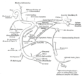 Plan of the facial and intermediate nerves and their communication with other nerves.
 -
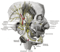
 The course and connections of the facial nerve in the temporal bone.
 -
 Upper part of medulla spinalis and hind- and mid-brains; posterior aspect, exposed in situ.
 -
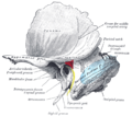 Left temporal bone showing surface markings for the tympanic antrum (red), transverse sinus (blue), and facial nerve (yellow).
 -
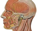 Head facial nerve branches
 -
 Facial nerve. Deep dissection.


See also
References
![]() This article incorporates text in the public domain from page 901 of the 20th edition of Gray's Anatomy (1918)
This article incorporates text in the public domain from page 901 of the 20th edition of Gray's Anatomy (1918)
- ^ a b "The Facial Nerve (CN VII)". TeachMeAnatomy. 2013-09-04. Retrieved 2018-05-04.
- ^ "Facial nerve | anatomy". Encyclopedia Britannica. Retrieved 2020-10-13.
- PMID 27040583.
- ^ "Parotidectomy with Facial Nerve Dissections | Iowa Head and Neck Protocols". medicine.uiowa.edu. Retrieved 2023-11-05.
- PMID 23766904.
- ISBN 9781451110326.
- ^ Singh V. Textbook of Clinical Neuroanatomy (2nd ed.). p. 104.
- PMID 24605304.
- ISBN 9781451190380.
- ISBN 9781437720020.
- S2CID 206906594.
- ISBN 91-44-37271-X
External links
| National | |
|---|---|
| Other | |