Fasciola gigantica
| Fasciola gigantica | |
|---|---|

| |
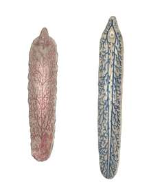
| Cobbold's drawings of dorsal (left) and ventral views of Fasciola gigantica | |
| Scientific classification | |
| Domain: | Eukaryota |
| Kingdom: | Animalia |
| Phylum: | Platyhelminthes |
| Class: | Trematoda |
| Order: | Plagiorchiida |
| Family: | Fasciolidae |
| Genus: | Fasciola |
| Species: | F. gigantica
|
| Binomial name | |
| Fasciola gigantica | |
Fasciola gigantica is a parasitic
. The infection is commonly called fasciolosis.The prevalence of F. gigantica often overlaps with that of Fasciola hepatica, and the two species are so closely related in terms of genetics, behaviour, and morphological and anatomical structures that distinguishing them is notoriously difficult.[2] Therefore, sophisticated molecular techniques are required to correctly identify and diagnose the infection.[3]
Distribution
Fasciola gigantica causes outbreaks in tropical areas of South Asia, Southeast Asia, and Africa. The geographical distribution of F. gigantica overlaps with F. hepatica in many African and Asian countries and sometimes in the same country, although in such cases, the ecological requirement of the flukes and their snail hosts are distinct. Infection is most prevalent in regions with intensive sheep and cattle production. In Egypt, F. gigantica has existed in domestic animals since the times of the pharaohs.[4]
Lifecycle
The lifecycle of F. gigantica is: Eggs (transported with feces) → egg hatch →
Intermediate hosts
As with other trematodes, Fasciola spp. develop in a
Definitive hosts
F. gigantica is a causative agent (together with F. hepatica) of fascioliasis in
The
Infection and pathogenicity
Infection with Fasciola spp. occurs when
Diagnosis
Despite the importance to differentiate between the infection by either fasciolid species, due to their distinct epidemiological, pathological, and control characteristics, unfortunately,
Treatment
Triclabendazole is the drug of choice in fasciolosis, as it is highly effective against both mature and immature flukes. Artemether has been demonstrated in vitro to be equally effective.[9] Though slightly less potent, artesunate is also useful in human fasciolosis.[10]
References
This article incorporates CC-BY-3.0 text from references.[4][11]
- ^ Cobbold, T. S. (1855). Description of a new trematode worm (Fasciola gigantica). The Edinburgh New Philosophical Journal, Exhibiting a View of the Progressive Discoveries and Improvements in the Sciences and the Arts. New Series, II, 262–267.
- PMID 21767436.
- ^ PMID 19769969.
- ^ PMID 19738348.

- PMID 21143890.
- PMID 16083061.
- PMID 22638917.
- PMID 20933335.
- PMID 19036519.
- PMID 18337331.
- ^ Onocha P. & Otunla E. (2008). "Biological activities of extracts of Pycnanthus angolensis (Welw.) Warb". African Journal of Traditional, Complementary and Alternative medicines, Abstracts of the world congress on medicinal and aromatic plants, Cape Town, November 2008. abstract Archived 2012-03-12 at the Wayback Machine
Further reading
- Le, T. H.; De, N. V.; Agatsuma, T.; Blair, D.; Vercruysse, J.; Dorny, P.; Nguyen, T. G. T.; McManus, D. P. (2006). "Molecular confirmation that Fasciola gigantica can undertake aberrant migrations in human hosts". Journal of Clinical Microbiology. 45 (2): 648–650. PMID 17135435.
- Meemon, K.; Grams, R.; Vichasri-Grams, S.; Hofmann, A.; Korge, G. N.; Viyanant, V.; Upatham, E. S.; Habe, S.; Sobhon, P. (2004). "Molecular cloning and analysis of stage and tissue-specific expression of cathepsin B encoding genes from Fasciola gigantica". Molecular and Biochemical Parasitology. 136 (1): 1–10. PMID 15138062.
- Spithill T. M.; Smooker P. M.; Copeman D. B. (1999). "Fasciola gigantica: epidemiology, control, immunology and molecular biology". In Dalton J. P. (ed.). Fasciolosis. Oxin, UK.: CABI Publishing. pp. 465–525.
