
Femoral vein
| Femoral vein | |
|---|---|
profunda femoris, great saphenous | |
| Drains to | external iliac vein |
| Artery | femoral artery |
| Identifiers | |
| Latin | vena femoralis |
| MeSH | D005268 |
| TA98 | A12.3.11.023 |
| TA2 | 5055 |
| FMA | 21185 |
| Anatomical terminology] | |
In the
Structure
The femoral vein bears valves which are mostly bicuspid and whose number is variable between individuals and often between left and right leg.[1]
Course

The femoral vein continues into the
The common femoral vein is the segment of the femoral vein between the branching point of the
Distal segment

In the past, the femoral vein was seen to follow the
Because of the widespread misunderstanding, and possible harmful results from the use of superficial femoral vein, a consensus was arrived at in 2001 during the World Congress of the International Union of Phlebology to change the name from superficial femoral vein simply to femoral vein.[13] This has been widely recognised and adopted though the use of superficial femoral vein still persists in some sources. Its use is actively discouraged.[14][15][16] It has been suggested that another term be used – the subsartorial vein.[17][18] A previous usage of subsartorial artery was published to avoid the name superficial femoral vein from being used.[19] As per the consensus of 2002, the superficial femoral artery was unchanged.[20]
Tributaries
The great saphenous vein, and the deep femoral vein are two large tributaries that drain into the femoral vein which then becomes known as the common femoral vein. Other smaller vein tributaries are the lateral and medial circumflex femoral veins.[21] These circumflex veins follow the lateral circumflex femoral artery, and the medial circumflex femoral artery.
Clinical significance
The femoral vein is a common site for a
The femoral vein is often used to place a
The practice of delivering
Additional images
-
 Position of femoral vein and artery in adductor canal
 -
 Structures surrounding right hip-joint.
 -
 Femoral sheath laid open to show its three compartments.
 -
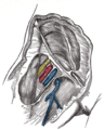
 Femoral vein


References
- ^ S2CID 52308003.
- ISBN 9780071222075.
- ^ Oh, Geon. "Femoral vein | Radiology Reference Article | Radiopaedia.org". Radiopaedia. Retrieved February 12, 2023.
- PMID 28260355.
- ^ Craig Hacking (August 26, 2019). "Common femoral vein". Radiopaedia.
- ISBN 9780323292177.
- S2CID 26014993.
- S2CID 8747016.
- PMID 12170230.)
{{cite journal}}: CS1 maint: multiple names: authors list (link - PMID 20445717.
- ISBN 9788131263617. Page 1072
- PMID 14595157.
- PMID 15874941. Retrieved February 12, 2023.
- S2CID 250044.
- S2CID 23215861.
- PMID 7563535.
- ^ Mikael Häggström (2019). "Subsartorial Vessels as Replacement Name for Superficial Femoral Vessels" (PDF). International Journal of Anatomy, Radiology and Surgery: AV01–AV02.
- ISBN 9788131263617. Page 1072
- ^ Antoine Micheau, MD. "Superficial femoral artery". IMAIOS. Retrieved December 22, 2022.
- S2CID 202165116.
- ^ "Femoral Vein: Anatomy & Function". Cleveland Clinic. Retrieved February 16, 2023.
- PMID 25598728.
- PMID 28844205.
- ISBN 978-0-323-03004-5, retrieved November 19, 2020
- ^ ISBN 978-0-443-10147-2, retrieved November 19, 2020
- ISBN 978-0-7020-6285-8
- PMID 15833116.
External links
- Anatomy figure: 12:05-01 at Human Anatomy Online, SUNY Downstate Medical Center - "Veins of the lower extremity shown in association with major landmarks."
- Cross section image: pelvis/pelvis-e12-15—Plastination Laboratory at the Medical University of Vienna