
Femur
| Femur | |
|---|---|
vastus intermedius | |
| Insertions | Gluteus maximus, gluteus medius, gluteus minimus, iliopsoas, lateral rotator group, adductors of the hip |
| Articulations | hip: acetabulum of pelvis superiorly knee: with the tibia and patella inferiorly |
| Identifiers | |
| Latin | os femoris, os longissimum |
| MeSH | D005269 |
| TA98 | A02.5.04.001 |
| TA2 | 1360 |
| FMA | 9611 |
| Anatomical terms of bone] | |
The femur (
The top of the femur fits into a socket in the pelvis called the hip joint, and the bottom of the femur connects to the shinbone (tibia) and kneecap (patella) to form the knee. In humans the femur is the largest and thickest bone in the body.
Structure
The femur is the only bone in the upper
In the condition genu valgum (knock knee) the femurs converge so much that the knees touch one another. The opposite extreme is genu varum (bow-leggedness). In the general population of people without either genu valgum or genu varum, the femoral-tibial angle is about 175 degrees.[3]
The femur is the largest and thickest bone in the human body. By some tested measures,[
The femur is categorised as a long bone and comprises a diaphysis (shaft or body) and two epiphyses (extremities) that articulate with adjacent bones in the hip and knee.[3]
Upper part
The
The
The transition area between the head and neck is quite rough due to attachment of muscles and the
A slight ridge is sometimes seen commencing about the middle of the intertrochanteric crest, and reaching vertically downward for about 5 cm. along the back part of the body: it is called the
About the junction of the upper one-third and lower two-thirds on the intertrochanteric crest is the quadrate tubercle located. The size of the tubercle varies and it is not always located on the intertrochanteric crest and that also adjacent areas can be part of the quadrate tubercle, such as the posterior surface of the greater trochanter or the neck of the femur. In a small anatomical study it was shown that the epiphyseal line passes directly through the quadrate tubercle.[5]
Body
.png)
The body of the femur (or shaft) is large, thick and almost cylindrical in form. It is a little broader above than in the center, broadest and somewhat flattened from before backward below. It is slightly arched, so as to be convex in front, and concave behind, where it is strengthened by a prominent longitudinal ridge, the linea aspera which diverges proximally and distal as the medial and lateral ridge. Proximally the lateral ridge of the linea aspera becomes the gluteal tuberosity while the medial ridge continues as the pectineal line. Besides the linea aspera the shaft has two other bordes; a lateral and medial border. These three bordes separates the shaft into three surfaces: One anterior, one medial and one lateral. Due to the vast musculature of the thigh the shaft can not be palpated.[3]
The third trochanter is a bony projection occasionally present on the proximal femur near the superior border of the gluteal tuberosity. When present, it is oblong, rounded, or conical in shape and sometimes continuous with the gluteal ridge.[6] A structure of minor importance in humans, the incidence of the third trochanter varies from 17–72% between ethnic groups and it is frequently reported as more common in females than in males.[7]
Lower part

The
Anteriorly, the condyles are slightly prominent and are separated by a smooth shallow articular depression called the patellar surface. Posteriorly, they project considerably and a deep notch, the
The articular surface of the lower end of the femur occupies the anterior, inferior, and posterior surfaces of the condyles. Its front part is named the patellar surface and articulates with the patella; it presents a median groove which extends downward to the intercondyloid fossa and two convexities, the lateral of which is broader, more prominent, and extends farther upward than the medial.[3]
Each condyle is surmounted by an elevation, the
Development
The femur develops from the limb buds as a result of interactions between the ectoderm and the underlying mesoderm; formation occurs roughly around the fourth week of development.[8]
By the sixth week of development, the first
Function
As the femur is the only bone in the thigh, it serves as an attachment point for all the muscles that exert their force over the hip and knee joints. Some
In cross-section, the thigh is divided up into three separate
Muscle attachments
 (seen from the front) |
 (seen from the back) |
| Muscle | Direction | Attachment[9] |
| Iliacus muscle | Insertion | Lesser trochanter |
| Psoas major muscle | Insertion | Lesser trochanter |
Gluteus maximus muscle |
Insertion | Gluteal tuberosity |
Gluteus medius muscle |
Insertion | Lateral surface of greater trochanter |
Gluteus minimus muscle |
Insertion | Forefront of greater trochanter |
| Piriformis muscle | Insertion | Superior boundary of greater trochanter |
Gemellus superior muscle |
Insertion | Upper edge of Obturator internus's tendon (indirectly greater trochanter )
|
| Obturator internus muscle | Insertion | Medial surface of greater trochanter |
Gemellus inferior muscle |
Insertion | Lower edge of Obturator internus's tendon (indirectly greater trochanter )
|
| Quadratus femoris muscle | Insertion | Intertrochanteric crest |
Obturator externus muscle |
Insertion | Trochanteric fossa |
| Pectineus muscle | Insertion | Pectineal line |
| Adductor longus muscle | Insertion | Medial ridge of linea aspera |
| Adductor brevis muscle | Insertion | Medial ridge of linea aspera |
| Adductor magnus muscle | Insertion | Medial ridge of linea aspera and the adductor tubercle |
| Vastus lateralis muscle | Origin | Greater trochanter and lateral ridge of linea aspera |
| Vastus intermedius muscle | Origin | Front and lateral surface of femur |
Vastus medialis muscle |
Origin | Distal part of intertrochanteric line and medial ridge of linea aspera |
| Short head of biceps femoris | Origin | Lateral ridge of linea aspera |
| Popliteus muscle | Origin | Under the lateral epicondyle |
Articularis genu muscle |
Origin | Lower 1/4 of anterior femur deep to vastus intermedius |
| Gastrocnemius muscle | Origin | Behind the adductor tubercle, over the lateral epicondyle and the popliteal facies |
| Plantaris muscle | Origin | Over the lateral condyle
|
Clinical significance
Fractures
A
Diversity among animals
-from-the-Genus-pone.0099929.g002.jpg)
In primitive tetrapods, the main points of muscle attachment along the femur are the internal trochanter and third trochanter, and a ridge along the ventral surface of the femoral shaft referred to as the adductor crest. The neck of the femur is generally minimal or absent in the most primitive forms, reflecting a simple attachment to the acetabulum. The greater trochanter was present in the extinct archosaurs, as well as in modern birds and mammals, being associated with the loss of the primitive sprawling gait. The lesser trochanter is a unique development of mammals, which lack both the internal and fourth trochanters. The adductor crest is also often absent in mammals or alternatively reduced to a series of creases along the surface of the bone.[10] Structures analogous to the third trochanter are present in mammals, including some primates.[7]
Some species of whales,[11] snakes, and other non-walking vertebrates have vestigial femurs. In some snakes the protruding end of a pelvic spur, a vestigial pelvis and femur remnant which is not connected to the rest of the skeleton, plays a role in mating. This role in mating is hypothesized to have possibly occurred in Basilosauridae, an extinct family of whales with well-defined femurs, lower legs and feet. Occasionally, the genes that code for longer extremities cause a modern whale to develop miniature legs (atavism).[12]
One of the earliest known vertebrates to have a femur is the
Viral metagenomics
A recent study revealed that bone is a much richer source of persistent DNA viruses than earlier perceived. Besides Parvovirus 19 and hepatitis B virus, ten additional ones were discovered, namely several members of the herpes- and polyomavirus families, as well as human papillomavirus 31 and torque teno virus. [13]
Invertebrates
In
Additional media
-
 3D image
 -
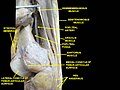 Muscles of thigh. Lateral view.
 -
 Muscles of thigh. Cross section.
 -
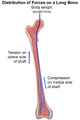 Distribution forces of the femur
 -
Femur Anatomy


References
- ^ "femora". Merriam-Webster.com Dictionary.
- ^ "femora". Dictionary.com Unabridged (Online). n.d.
- ^ ISBN 978-87-628-0307-7.
- PMID 2252082.)
{{cite journal}}: CS1 maint: multiple names: authors list (link - PMID 17104699.
- PMID 3870721.
- ^ a b
Bolanowski, Wojciech; Śmiszkiewicz-Skwarska, Alicja; Polguj, Michał; Jędrzejewski, Kazimierz S (2005). "The occurrence of the third trochanter and its correlation to certain anthropometric parameters of the human femur" (PDF). Folia Morphol. 64 (3): 168–175. PMID 16228951.
- ^ Gilbert, Scott F. "Developmental Biology". 9th ed., 2010
- ISBN 978-87-628-0307-7.
- ISBN 978-0-03-910284-5.
- PMID 17231384.
- S2CID 8448387.
- S2CID 220582800.