
File:6nb6 prefusion 6m3w postfusion spike.png
Original file (768 × 1,536 pixels, file size: 382 KB, MIME type: image/png)
| This is a file from the Wikimedia Commons. Information from its description page there is shown below. Commons is a freely licensed media file repository. You can help. |
Summary
| Description |
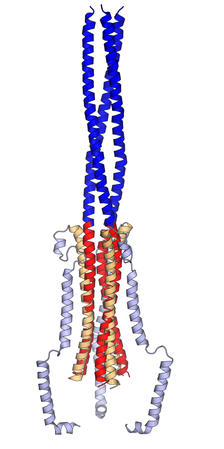
Comparison of the pre-fusion (orange, light blue) and post-fusion (red, dark blue) conformations of the SARS-CoV spike protein trimer. In the pre-fusion conformation, the central helix (orange) and heptad repeat 1 (HR1, light blue) are folded back on each other in an antiparallel orientation. In the post-fusion conformation, the central helix (red) and the HR1 sequence (dark blue) reorganize to form an extended trimeric coiled coil. The viral membrane is at the bottom and the host cell membrane at the top. Only key portions of the S2 subunit are shown. Rendered using PyMol from cryo-electron microscopy structures PDB: 6NB6 (pre-fusion) and PDB: 6M3W (post-fusion) superposed using the central helix sequences, inspired by Figs 1 and 2 from Fan 2020. 6NB6: Unexpected Receptor Functional Mimicry Elucidates Activation of Coronavirus Fusion. Walls, A.C., Xiong, X., Park, Y.J., Tortorici, M.A., Snijder, J., Quispe, J., Cameroni, E., Gopal, R., Dai, M., Lanzavecchia, A., Zambon, M., Rey, F.A., Corti, D., Veesler, D. (2019) Cell 176: 1026-1039.e15 PubMed: 30712865 DOI: 10.1016/j.cell.2018.12.028 6M3W: Cryo-EM analysis of the post-fusion structure of the SARS-CoV spike glycoprotein. Fan, X., Cao, D., Kong, L., Zhang, X. (2020) Nat Commun 11: 3618-3618 PubMed: 32681106 DOI: 10.1038/s41467-020-17371-6 |
| Date | |
| Source | Own work |
| Author | Opabinia regalis |
Licensing
- You are free:
- to share – to copy, distribute and transmit the work
- to remix – to adapt the work
- Under the following conditions:
- attribution – You must give appropriate credit, provide a link to the license, and indicate if changes were made. You may do so in any reasonable manner, but not in any way that suggests the licensor endorses you or your use.
- share alike – If you remix, transform, or build upon the material, you must distribute your contributions under the same or compatible license as the original.

|
Permission is granted to copy, distribute and/or modify this document under the terms of the GNU Free Documentation License, Version 1.2 or any later version published by the Free Software Foundation; with no Invariant Sections, no Front-Cover Texts, and no Back-Cover Texts. A copy of the license is included in the section entitled GNU Free Documentation License. |
File history
Click on a date/time to view the file as it appeared at that time.
| Date/Time | Thumbnail | Dimensions | User | Comment | |
|---|---|---|---|---|---|
| current | 07:02, 13 September 2021 | | 768 × 1,536 (382 KB) | Opabinia regalis | {{Information |Description=Comparison of the pre-fusion (orange, light blue) and post-fusion (red, dark blue) conformations of the SARS-CoV spike protein trimer. In the pre-fusion conformation, the central helix (orange) and heptad repeat 1 (HR1, light blue) are folded back on each other in an antiparallel orientation. In the post-fusion conformation, the central helix (red) and the HR1 sequence (dark blue) reorganize to form an extended trimeric coiled coil. The viral membrane is at the bott... |
File usage
Global file usage
The following other wikis use this file:
- Usage on ar.wikipedia.org
- Usage on ca.wikipedia.org
- Usage on de.wikipedia.org
- Usage on es.wikipedia.org