
Search results
There is a page named "File:HypothalamicNuclei.PNG" on Wikipedia
 Hypothalamic Nuclei Symbols: AC: anterior commissure PO: preoptic nucleus SC: suprachiasmatic nucleus OC: optic chiasma TC: tuber cinereum AP: anterior...(769 × 603 (239 KB)) - 21:09, 19 February 2022
 BY-SA 4.0 Creative Commons Attribution-Share Alike 4.0 truetrue English Coronal view of the approximate location of the hypothalamic nuclei and tanycytes...(736 × 546 (534 KB)) - 03:46, 15 September 2020
 0 Creative Commons Attribution-Share Alike 4.0 truetrue English hypothalamic nuclei and the distribution of tanycytes over the wall of the third ventricle...(1,278 × 547 (774 KB)) - 02:03, 30 December 2023
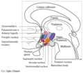 DescriptionHypothalamic nuclei.png English: Hypothalamic nuclei. Linkage between the central nervous system and the pituitary gland. Date 28 July 2013...(463 × 410 (187 KB)) - 04:37, 17 September 2020
 DescriptionSchematic representation of the hypothalamic nuclei.png English: A schematic representation of the hypothalamic nuclei and the distribution of tanycytes...(1,276 × 546 (539 KB)) - 11:10, 6 September 2020
 diferentes núcleos que forman el hipotálamo. Date 18 November 2019 Source WIKIMEDIA COMMONS File:Hypothalamic nuclei.png Author Zsuzsanna Suba (modificado)...(926 × 820 (188 KB)) - 08:12, 30 October 2020
 DescriptionKey hypothalamic nuclei and other areas involved in glucose homeostasis.png English: Key hypothalamic nuclei and other areas involved in glucose...(1,944 × 1,084 (355 KB)) - 19:49, 5 September 2020
 pathways of the suprachiasmatic nuclei (SCN).png English: Input and output pathways to/from the suprachiasmatic nuclei (SCN). The photic input pathways...(1,296 × 669 (398 KB)) - 14:35, 16 February 2023
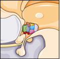 DescriptionServier Medical Art Hypothalamus 3.png English: Hypothalamic nuclei. Date 1 August 2012 Source https://smart.servier.com/?s=Suprachiasmatic+nucleus...(259 × 256 (30 KB)) - 16:25, 12 September 2020
 DescriptionServier Medical Art Hypothalamus 2.png English: Hypothalamic nuclei. Date 1 August 2012 Source https://smart.servier.com/?s=Suprachiasmatic+nucleus...(320 × 320 (37 KB)) - 14:07, 1 October 2020
 DescriptionServier Medical Art Hypothalamus 1.png English: Hypothalamic nuclei. Date 1 August 2012 Source https://smart.servier.com/?s=Suprachiasmatic+nucleus...(256 × 196 (24 KB)) - 14:07, 1 October 2020
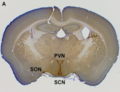 26 April 2012 Source DLK1 Is a Somato-Dendritic Protein Expressed in Hypothalamic Arginine-Vasopressin and Oxytocin Neurons. PLoS ONE 7(4): e36134. doi:10...(621 × 474 (363 KB)) - 01:28, 19 February 2024
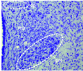 Barra de escala: 50 µm. Date 7 August 2013 Source Bilateral Descending Hypothalamic Projections to the Spinal Trigeminal Nucleus Caudalis in Rats. PLoS ONE...(744 × 636 (925 KB)) - 22:19, 16 November 2022
 DescriptionNucleo Arcuato.png Español: Figura 9. MCT2 se localiza en neuronas AgRP y POMC del núcleo arqueado (AN). b) Reconstrucción anteroposterior...(2,988 × 686 (2.24 MB)) - 08:21, 4 January 2022
 DescriptionNucleo Arcuato AN.png Español: Figura 9. MCT2 se localiza en neuronas AgRP y POMC del núcleo arqueado. (b) Reconstrucción anteroposterior del...(576 × 670 (288 KB)) - 14:34, 31 December 2021
 DescriptionNúcleo Arcuato lateral.png Español: Figura 11. MCT2 se localiza en neuronas anorexigénicas de Núcleo Arcuato (AN). a – c) Imágenes de bajo...(2,826 × 844 (2.04 MB)) - 13:00, 17 May 2022
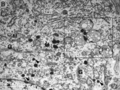 DescriptionMagnocel Neurosecre Hipotal.png Español: Fig 2. Descripción ultraestructural de las neuronas magnocelulares el núcleo supraóptico (SON) y sus...(1,031 × 776 (772 KB)) - 04:02, 8 October 2020
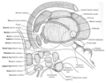 DescriptionLawrence 1960 22.3.png Anatomical illustration of the human nervous system from the 1960 book A functional approach to neuroanatomy Source...(2,304 × 1,804 (1.24 MB)) - 12:01, 19 January 2020
 DescriptionNucleo Arcuato Tanicitos tipos.png Español: Figura 7. MCT2 se localiza en el núcleo arqueado. (b) Sección del cerebro frontal de rata usando...(608 × 714 (599 KB)) - 14:28, 31 December 2021
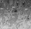 DescriptionNúcleo Supraopt Capilares.png Español: Fig 3. Poblaciones celulares y vasculatura del núcleo supraóptico SON en la zona somática. B) El núcleo...(1,035 × 968 (748 KB)) - 04:04, 18 November 2020


.png)


