
Search results
There is a page named "File:Kidney section.jpg" on Wikipedia
 DescriptionKidney section.jpg Section of kidney Plate InfoField 1127 Date before 1858 date QS:P,+1858-00-00T00:00:00Z/7,P1326,+1858-00-00T00:00:00Z/9 Source...(208 × 350 (37 KB)) - 19:09, 6 November 2021
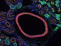 0CC BY 4.0 Creative Commons Attribution 4.0 truetrue English Mouse Kidney Section Wikimedia username: Erin Rod URL: https://commons.wikimedia.org/wiki/user:Erin_Rod...(1,920 × 1,452 (255 KB)) - 06:06, 18 November 2022
 0CC BY 4.0 Creative Commons Attribution 4.0 truetrue English Mouse Kidney Section 3 Wikimedia username: Erin Rod author name string: Erin Rod URL: https://commons...(1,920 × 1,452 (382 KB)) - 06:06, 18 November 2022
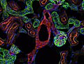 0CC BY 4.0 Creative Commons Attribution 4.0 truetrue English Mouse Kidney Section 2 author name string: Erin Rod Wikimedia username: Erin Rod URL: https://commons...(1,920 × 1,452 (290 KB)) - 06:06, 18 November 2022
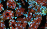 DescriptionMouse Kidney (23725924684).jpg Mouse kidney section, DAPI nuclei, Alexa Fluor 488 Wheat Germ Agglutinin, Alexa Fluor 568 Phalloidin actin....(1,916 × 1,210 (993 KB)) - 20:09, 16 December 2023
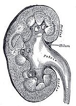 Section of kidney, from Gray's Anatomy 1918....(208 × 290 (17 KB)) - 07:14, 5 February 2012
 http://arachnophiliac.info Wikicommon's Image:Common snail.jpg English A diagram showing a split-sectioned drawing of a snail, with numbers from 1 to 24 pointing...(700 × 400 (211 KB)) - 09:40, 28 March 2024
 Attribution 4.0 truetrue English Author's drawing a cross-section of a dolphin and its reniculate kidney Russian Авторский рисунок поперечного сечения дельфина...(1,200 × 3,970 (440 KB)) - 04:03, 24 October 2023
 2: Cortex 3: Medulla 4: Perirenal fat 5: Capsule 6: Ureter 7: Pelvis of kidney 8: Renal vessels 9: Hilus 10: Calyx https://creativecommons.org/publicdomain/mark/1...(319 × 260 (43 KB)) - 11:29, 10 September 2022
 English Image of a close up nephron and its place in the kidney....(2,044 × 1,890 (1.17 MB)) - 06:56, 31 October 2022
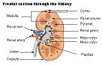 seer.cancer.gov/module_anatomy/unit11_2_uri_comp1_kidney.html Language neutral version:Image:Kidney_structure_neutral.png https://creativecommons...(400 × 256 (30 KB)) - 16:21, 16 August 2024
 English Constance Tom Noguchi, Ph.D., section chief at National Institute of Diabetes and Digestive and Kidney Diseases, National Institutes of Health...(300 × 300 (30 KB)) - 06:19, 12 April 2024
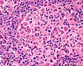 English Tetracapsuloides bryosalmonae parasites in rainbow trout kidney. Tissue section stained with haematoxylin and eosin. Photo courtesy of Cefas. URL:...(1,232 × 972 (1.04 MB)) - 16:17, 5 December 2023
 is an ultrasonography scan of the longitudinal view of the normal left kidney. Prof Dr. Abdul Kareem.M.M. object has role: photographer Wikimedia username:...(800 × 600 (50 KB)) - 03:10, 20 September 2020
 Histopath section micrograph of a FIP-infected kidney showing characteristic inflammatory response. Original caption: "Kidney, the inflammatory cells...(350 × 246 (66 KB)) - 18:01, 24 September 2020
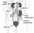 {{Information |Description=Kidney location after transplantation. |Source=[http://kidney.niddk.nih.gov/kudiseases/pubs/transplant http://kidney.niddk.nih...(598 × 583 (42 KB)) - 11:47, 17 August 2024
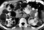 polycystic kidney disease. Note extensive bilateral renal and pancreatic cysts. This pattern can mimic autosomal dominant polycystic kidney disease,including...(468 × 321 (73 KB)) - 05:11, 18 August 2024
 Attribution 3.0 truetrue English Labeled illustration of the anatomy of human kidneys French Rein author name string: BruceBlaus Wikimedia username: BruceBlaus...(1,600 × 1,200 (1.17 MB)) - 15:24, 4 August 2024
 171 75 137 116 623 448 Right kidney with enlarged pelvis 389 70 115 85 623 448 Left kidney English URL: https://commons.wikimedia.org/wiki/user:Nevit...(623 × 448 (65 KB)) - 07:16, 1 January 2023
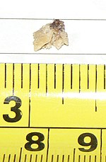 following user names refer to en.wikipedia. 2007-03-05 00:26 Melensdad 800×1224×8 (147839 bytes) Kidney Stone that passed naturally, albeit painfully. English...(800 × 1,224 (144 KB)) - 13:44, 25 April 2022


.jpg)


