
Fish fin
Fins are moving
.Fish fins are distinctive anatomical features with varying structures among different
Fins at different locations on the fish serve different purposes, and are divided into two groups: the
Types of fins
Fins can either be paired or unpaired. The pectoral and pelvic fins are paired, whereas the dorsal, anal and caudal fins are unpaired and situated along the midline of the body. For every type of fin, there are a number of fish
| Pectoral fins (Arm fins) |

|
The paired pectoral fins are located on each side, usually kept folded just behind the operculum, and are homologous to the forelimbs of quadrupedal tetrapods or the upper limbs of bipedal tetrapods.
|
|---|---|---|
| Pelvic / Ventral fins (Belly fins) |

|
The paired pelvic or ventral fins are the belly fins (from Latin venter 'belly') are typically located ventrally below and behind the pectoral fins, although in many fish families they may be positioned in front of the pectoral fins (e.g. cods). They are homologous to the hindlimbs of quadrupedal tetrapods or the lower limbs of bipedal tetrapods.
The pelvic fin assists the fish in going up or down through the water, turning sharply, and stopping quickly.
|
| Dorsal fin (Spinal fins) |
 Dorsal fin of a shark
|
 The dorsal fins are located on the back. A fish can have up to three dorsal fins. The dorsal fins serve to protect the fish against rolling, and assist it in sudden turns and stops.
|
| Anal/cloacal fin | 
|
The anal/cloacal fin is located on the ventral surface behind the anus/cloaca.
|
| Adipose fin |  Adipose fin of a trout
|
The adipose fin is a soft, fleshy fin found on the back behind the dorsal fin and just forward of the caudal fin. It is absent in many fish families, but found in nine of the 31 characids and catfish .
The function of the adipose fin is something of a mystery. It is frequently clipped off to mark hatchery-raised fish, though data from 2005 showed that trout with their adipose fin removed have an 8% higher tailbeat frequency.[4][5] Additional information released in 2011 has suggested that the fin may be vital for the detection of, and response to, stimuli such as touch, sound and changes in pressure. Canadian researchers identified a neural network in the fin, indicating that it likely has a sensory function, but are still not sure exactly what the consequences of removing it are.[6][7] A comparative study in 2013 indicates the adipose fin can develop in two different ways. One is the salmoniform-type way, where the adipose fin develops from the larval-fin fold at the same time and in the same direct manner as the other median fins. The other is the characiform-type way, where the adipose fin develops late after the larval-fin fold has diminished and the other median fins have developed. They claim the existence of the characiform-type of development suggests the adipose fin is not "just a larval fin fold remainder" and is inconsistent with the view that the adipose fin lacks function.[3] Research published in 2014 indicates that the adipose fin has evolved repeatedly in separate lineages.[8] |
| Caudal fin (Tail fin) |
  Heterocercal caudal fin (A)  Homocercal caudal fin (C)
|
The caudal fin is the tail fin (from the Latin cauda meaning tail), located at the end of the caudal peduncle. It is used for propulsion in most vertebrae of the axial skeleton and pterygiophores (radials). Depending on the relationship with the axial skeleton, four types of caudal fins (A-D) are distinguished:
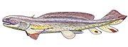
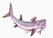
(A) - Heterocercal means the vertebrae extend into the upper lobe of the tail, often making it longer than the lower lobe (as in stem Actinopterygii, and sturgeons and paddlefish). However, the external shape of heterocercal tail fins can also appear symmetric (e.g. †Birgeria, †Bobasatrania ). Heterocercal is the opposite of hypocercal
(B) - Protocercal means the vertebrae extend to the tip of the tail and the tail is symmetrical but not expanded (as in lancelets )
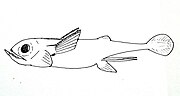
(C) - Homocercal where the fin usually appears superficially symmetric but in fact the vertebrae extend for a very short distance into the upper lobe of the fin. Homocercal caudal fins can, however, also appear asymmetric (e.g. blue flying fish). Most modern fishes (teleosts) have a homocercal tail. These come in a variety of shapes, and can appear:
(D) - Diphycercal means the vertebrae extend to the tip of the tail and the tail is symmetrical and expanded (as in the Palaeozoic fishes had a diphycercal heterocercal tail.[11]
|
| Caudal keel Finlets |
_diagram_cropped.GIF)
|
Some types of fast-swimming fish have a horizontal caudal keel just forward of the tail fin. Much like the keel of a ship, this is a lateral ridge on the caudal peduncle, usually composed of scutes (see below), that provides stability and support to the caudal fin. There may be a single paired keel, one on each side, or two pairs above and below.
Finlets are small fins, generally behind the dorsal and anal fins (in sauries , they are rayless, non-retractable, and found between the last dorsal and/or anal fin and the caudal fin.
|
Bony fishes
Bony fishes are divided into
Bony fish have fin spines called
Lobe-fins

The coelacanth is one type of living lobe-finned fish. Both extant members of this group, the West Indian Ocean coelacanth (Latimeria chalumnae) and the Indonesian coelacanth (Latimeria menadoensis), are found in the genus Latimeria. Coelacanths are thought to have evolved roughly into their current form about 408 million years ago, during the early Devonian.[18]
Locomotion of the coelacanths is unique to their kind. To move around, coelacanths most commonly take advantage of up or downwellings of the current and drift. They use their paired fins to stabilize their movement through the water. While on the ocean floor their paired fins are not used for any kind of movement. Coelacanths can create thrust for quick starts by using their caudal fins. Due to the high number of fins they possess, coelacanths have high maneuverability and can orient their bodies in almost any direction in the water. They have been seen doing headstands and swimming belly up. It is thought that their rostral organ helps give the coelacanth electroperception, which aids in their movement around obstacles.[19]
Fin arrangement and body shape is relatively conservative in lobe-finned fishes. However, there are a few examples from the
Diversity of fins in lobe-finned fishes
-
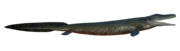 †Tiktaalik roseae
 -
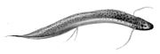 Spotted lungfishProtopterus dolloi
 -
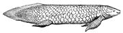 Queensland lungfishNeoceratodus forsteri
 -

-

-

-

-

-
 West Indian Ocean coelacanthLatimeria chalumnae
 -
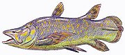 †Mawsonia gigas
_by_ObsidianSoul_02_flipped.png)


_(cropped).jpg)

.jpg)
Ray-fins

Spines have a variety of uses. In catfish, they are used as a form of defense; many catfish have the ability to lock their spines outwards. Triggerfish also use spines to lock themselves in crevices to prevent them being pulled out.
Lepidotrichia are usually composed of
Diversity of fins in ray-finned fishes
-
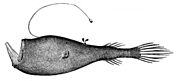
 Fanfin angler Caulophryne jordani
 -
 Pancake batfishHalieutichthys aculeatus
 -
 Slender sunfishRanzania laevis
 -
 Fanfish Pteraclis carolinus
 -
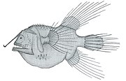
 Diaphanous hatchetfish Sternoptyx diaphana
 -
 Silver roughyHoplostethus mediterraneus
 -
 Crested flounder Lophonectes gallus
 -
 Jack-knifefish Equetus lanceolatus
 -
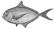 Atlantic pomfretBrama brama
 -
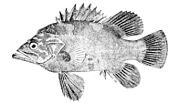 Atlantic wreckfishPolyprion americanus
 -
 Stellate pufferfish Arothron stellatus
 -
 Stargazing seadevil Ceratias uranoscopus
 -
 Ridgehead Poromitra unicornis
 -
 Tropical two-wing flyingfishExocoetus evolans
 -
 Cusk-eel Benthocometes robustus
 -
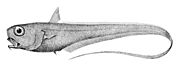 RattailTrachonurus sulcatus
 -
 Tripod fish Bathypterois grallator
 -
 Giant oarfish Regalecus glesne
 -
 Shortbill spearfish Tetrapturus angustirostris
 -
 Ghost knifefish Sternarchorhynchus oxyrhynchus
 -
 Remora Remora brachyptera
 -
 Blue-dashed rockskipperBlenniella periophthalmus
 -
 Nile bichirPolypterus bichir
 -
 Coastal cutthroat trout Oncorhynchus clarkii
 -
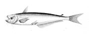 African butter catfish Schilbe mystus
 -
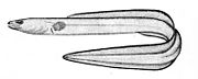 Conger eelLeptocephalus conger


.gif)


.gif)


.gif)
.gif)


Cartilaginous fishes

Shark fin skeletons are elongated and supported with soft and unsegmented rays named ceratotrichia, filaments of elastic protein resembling the horny keratin in hair and feathers.[23] Originally the pectoral and pelvic girdles, which do not contain any dermal elements, did not connect. In later forms, each pair of fins became ventrally connected in the middle when scapulocoracoid and puboischiadic bars evolved. In rays, the pectoral fins have connected to the head and are very flexible. One of the primary characteristics present in most sharks is the heterocercal tail, which aids in locomotion.[24] Most sharks have eight fins. Sharks can only drift away from objects directly in front of them because their fins do not allow them to move in the tail-first direction.[25]
Unlike modern cartilaginous fish, members of
As with most fish, the tails of sharks provide thrust, making speed and acceleration dependent on tail shape.
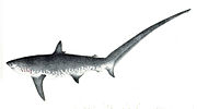
Tiger sharks have a large upper lobe, which allows for slow cruising and sudden bursts of speed. The tiger shark must be able to twist and turn in the water easily when hunting to support its varied diet, whereas the porbeagle shark, which hunts schooling fish such as mackerel and herring, has a large lower lobe to help it keep pace with its fast-swimming prey.[13] Other tail adaptations help sharks catch prey more directly, such as the thresher shark's usage of its powerful, elongated upper lobe to stun fish and squid.
On the other hand, rays rely on their enlarged pectoral fins for propulsion. Similarly enlarged pectoral fins can be found in the
)Diversity of fins in cartilaginous fishes
-
 Small-spotted catsharkScyliorhinus canicula
 -
 Great white sharkCarcharodon carcharias
 -
 Common thresherAlopias vulpinus
 -
 Largetooth sawfishPristis perotteti
 -
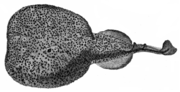 Marbled electric rayTorpedo marmorata
 -
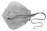 Bennett's stingrayHemitrygon bennettii
 -
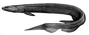 Frilled sharkChlamydoselachus anguineus
 -
-
 The †Stethacanthus productus
 -
 The †Wodnika striatula
 -
 Theelasmobranch †Bandringa rayi
 -
 The †Iniopterygiformes †Sibyrhynchus denisoni (Holocephali)
 -
 Cuban chimaeraChimaera cubana
 -
 American elephantfishCallorhinchus callorhynchus

.png)


Shark finning
According to the Humane Society International, approximately 100 million sharks are killed each year for their fins, in an act known as shark finning.[31] After the fins are cut off, the mutilated sharks are thrown back in the water and left to die.
In some countries of Asia, shark fins are a culinary delicacy, such as shark fin soup.[32] Currently, international concerns over the sustainability and welfare of sharks have impacted consumption and availability of shark fin soup worldwide.[33] Shark finning is prohibited in many countries.
Fin functions
Generating thrust

Cavitation occurs when negative pressure causes bubbles (cavities) to form in a liquid, which then promptly and violently collapse. It can cause significant damage and wear.[35] Cavitation damage can occur to the tail fins of powerful swimming marine animals, such as dolphins and tuna. Cavitation is more likely to occur near the surface of the ocean, where the ambient water pressure is relatively low. Even if they have the power to swim faster, dolphins may have to restrict their speed because collapsing cavitation bubbles on their tail are too painful.[36] Cavitation also slows tuna, but for a different reason. Unlike dolphins, these fish do not feel the bubbles, because they have bony fins without nerve endings. Nevertheless, they cannot swim faster because the cavitation bubbles create a vapor film around their fins that limits their speed. Lesions have been found on tuna that are consistent with cavitation damage.[36]
Fish use multiple fins, so it is possible that a given fin can have a hydrodynamic interaction with another fin. In particular, the fins immediately upstream of the caudal (tail) fin may be proximate fins that can directly affect the flow dynamics at the caudal fin. In 2011, researchers using volumetric imaging techniques were able to generate "the first instantaneous three-dimensional views of wake structures as they are produced by freely swimming fishes". They found that "continuous tail beats resulted in the formation of a linked chain of vortex rings" and that "the dorsal and anal fin wakes are rapidly entrained by the caudal fin wake, approximately within the timeframe of a subsequent tail beat".[40]
Controlling motion
Once motion has been established, the motion itself can be controlled with the use of other fins.[34][41]
The bodies of
Reproduction


Male
Gonopodia are found on the males of some species in the
Similar organs with similar characteristics are found in other fishes, for example the andropodium in the Hemirhamphodon or in the Goodeidae[51] or the gonopodium in the Middle Triassic †Saurichthys, the oldest known example of viviparity in a ray-finned fish.[52]
Other functions
Other uses of fins include walking and perching on the sea floor, gliding over water, cooling of body temperature, stunning of prey, display (scaring of predators, courtship), defence (venomous fin spines, locking between corals), luring of prey, and attachment structures.
The Indo-Pacific sailfish has a prominent dorsal fin. Like scombroids and other billfish, they streamline themselves by retracting their dorsal fins into a groove in their body when they swim.[55] The huge dorsal fin, or sail, of the sailfish is kept retracted most of the time. Sailfish raise them if they want to herd a school of small fish, and also after periods of high activity, presumably to cool down.[55][56]
_in_Waikiki_Aquarium.JPG)


The oriental flying gurnard has large pectoral fins which it normally holds against its body, and expands when threatened to scare predators. Despite its name, it is a demersal fish, not a flying fish, and uses its pelvic fins to walk along the bottom of the ocean.[58][59]
Fins can have an adaptive significance as sexual ornaments. During courtship, the female cichlid, Pelvicachromis taeniatus, displays a large and visually arresting purple pelvic fin. "The researchers found that males clearly preferred females with a larger pelvic fin and that pelvic fins grew in a more disproportionate way than other fins on female fish."[60][61]
Evolution
Evolution of paired fins
There are two prevailing hypotheses that have been historically debated as models for the evolution of paired fins in fish: the gill arch theory and the lateral fin-fold theory. The former, commonly referred to as the “Gegenbaur hypothesis,” was posited in 1870 and proposes that the “paired fins are derived from gill structures”.[63] This fell out of popularity in favor of the lateral fin-fold theory, first suggested in 1877, which proposes that paired fins budded from longitudinal, lateral folds along the epidermis just behind the gills.[64] There is weak support for both hypotheses in the fossil record and in embryology.[65] However, recent insights from developmental patterning have prompted reconsideration of both theories in order to better elucidate the origins of paired fins.
Classical theories
Carl Gegenbaur's concept of the “Archipterygium” was introduced in 1876.[66] It was described as a gill ray, or “joined cartilaginous stem,” that extended from the gill arch. Additional rays arose from along the arch and from the central gill ray. Gegenbaur suggested a model of transformative homology – that all vertebrate paired fins and limbs were transformations of the Archipterygium. Based on this theory, paired appendages such as pectoral and pelvic fins would have differentiated from the branchial arches and migrated posteriorly. However, there has been limited support for this hypothesis in the fossil record both morphologically and phylogenically.[65] In addition, there was little to no evidence of an anterior-posterior migration of pelvic fins.[67] Such shortcomings of the gill-arch theory led to its early demise in favor of the lateral fin-fold theory proposed by St. George Jackson Mivart, Francis Balfour, and James Kingsley Thacher.
The lateral fin-fold theory hypothesized that paired fins developed from lateral folds along the body wall of the fish.[64] Just as segmentation and budding of the median fin fold gave rise to the median fins, a similar mechanism of fin bud segmentation and elongation from a lateral fin fold was proposed to have given rise to the paired pectoral and pelvic fins. However, there was little evidence of a lateral fold-to-fin transition in the fossil record.[68] In addition, it was later demonstrated phylogenically that pectoral and pelvic fins arise from distinct evolutionary and mechanistic origins.[65]
Evolutionary developmental biology
Recent studies in the ontogeny and evolution of paired appendages have compared finless vertebrates – such as lampreys – with chondricthyes, the most basal living vertebrate with paired fins.[69] In 2006, researchers found that the same genetic programming involved in the segmentation and development of median fins was found in the development of paired appendages in catsharks.[70] Although these findings do not directly support the lateral fin-fold hypothesis, the original concept of a shared median-paired fin evolutionary developmental mechanism remains relevant.
A similar renovation of an old theory may be found in the developmental programming of chondricthyan gill arches and paired appendages. In 2009, researchers at the University of Chicago demonstrated that there are shared molecular patterning mechanisms in the early development of the chondricthyan gill arch and paired fins.[71] Findings such as these have prompted reconsideration of the once-debunked gill-arch theory.[68]
From fins to limbs
Fish are the ancestors of all mammals, reptiles, birds and amphibians.[72] In particular, terrestrial tetrapods (four-legged animals) evolved from fish and made their first forays onto land 400 million years ago.[73] They used paired pectoral and pelvic fins for locomotion. The pectoral fins developed into forelegs (arms in the case of humans) and the pelvic fins developed into hind legs.[74] Much of the genetic machinery that builds a walking limb in a tetrapod is already present in the swimming fin of a fish.[75][76]
Aristotle recognised the distinction between
homologous structures, and made the following prophetic comparison: "Birds in a way resemble fishes. For birds have their wings in the upper part of their bodies and fishes have two fins in the front part of their bodies. Birds have feet on their underpart and most fishes have a second pair of fins in their under-part and near their front fins."
– Aristotle, De incessu animalium[77]

In 2011, researchers at Monash University in Australia used primitive but still living lungfish "to trace the evolution of pelvic fin muscles to find out how the load-bearing hind limbs of the tetrapods evolved."[78][79] Further research at the University of Chicago found bottom-walking lungfishes had already evolved characteristics of the walking gaits of terrestrial tetrapods.[80][81]
In a classic example of convergent evolution, the pectoral limbs of pterosaurs, birds and bats further evolved along independent paths into flying wings. Even with flying wings there are many similarities with walking legs, and core aspects of the genetic blueprint of the pectoral fin have been retained.[82][83]
The first mammals appeared during the

"This sea-going reptile with terrestrial ancestors converged so strongly on fishes that it actually evolved a dorsal fin and tail fin for improved aquatic locomotion. These structures are all the more remarkable because they evolved from nothing — the ancestral terrestrial reptile had no hump on its back or blade on its tail to serve as a precursor."[90]
The biologist Stephen Jay Gould said the ichthyosaur was his favorite example of convergent evolution.[91]
Fins or flippers of varying forms and at varying locations (limbs, body, tail) have also evolved in a number of other tetrapod groups, including diving birds such as penguins (modified from wings), sea turtles (forelimbs modified into flippers), mosasaurs (limbs modified into flippers), and sea snakes (vertically expanded, flattened tail fin).
Robotic fins
| External videos | |
|---|---|
The use of fins for
The AquaPenguin, developed by Festo of Germany, copies the streamlined shape and propulsion by front flippers of penguins.[98][99] Festo also developed AquaRay,[100] AquaJelly[101] and AiraCuda,[102] respectively emulating the locomotion of manta rays, jellyfish and barracuda.
In 2004,
Robotic fish offer some research advantages, such as the ability to examine an individual part of a fish design in isolation from the rest of the fish. However, this risks oversimplifying the biology so key aspects of the animal design are overlooked. Robotic fish also allow researchers to vary a single parameter, such as flexibility or a specific motion control. Researchers can directly measure forces, which is not easy to do in live fish. "Robotic devices also facilitate three-dimensional kinematic studies and correlated hydrodynamic analyses, as the location of the locomotor surface can be known accurately. And, individual components of a natural motion (such as outstroke vs. instroke of a flapping appendage) can be programmed separately, which is certainly difficult to achieve when working with a live animal."[105]
See also
- Cephalopod fin
- Fin and flipper locomotion
- Fish locomotion
- Polydactyly in early tetrapods
- RoboTuna
- Shark fin soup
- Tradeoffs for locomotion in air and water
- Undulatory locomotion
References
Citations
- PMID 20154199.
- ^ Gene Helfman, Bruce Collette, Douglas Facey, & Brian Bowen. (2009) The Diversity of Fishes: biology, evolution, and ecology. John Wiley & Sons.
- ^ ISSN 1860-0743.
- .
- doi:10.1139/Z04-069.
- ^ Temple, Nicola (18 July 2011). "Removal of trout, salmon fin touches a nerve". Cosmos. Archived from the original on 12 January 2014.
- PMID 21733904.
- PMID 24598422.
- ISBN 978-0226870137. Retrieved 18 October 2018.
- ^ S2CID 84992418.
- ^ von Zittel KA, Woodward AS and Schlosser M (1932) Text-book of Paleontology Volume 2, Macmillan and Company. Page 13.
- S2CID 87234436.
- ^ OCLC 28965588.
- ^ a b "Osteichthyes - Bony Fish". Wildlife Journal Junior. New Hampshire PBS. 2023. Retrieved 31 March 2024.
- ^ Speer, B.R. (29 May 2000). "Introduction to the Dipnoi - the lungfish". University of California Museum of Paleontology. Retrieved 31 March 2024.
- PMID 26908371. Art. No. 21571.
- ^ Clack, J. A. (2002) Gaining Ground. Indiana University
- PMID 17148426.
- S2CID 4353395.
- PMID 28082606.
- ^ .
- S2CID 205221027.
- ^ Hamlett 1999, p. 528.
- ^ Function of the heterocercal tail in sharks: quantitative wake dynamics during steady horizontal swimming and vertical maneuvering - The Journal of Experimental Biology 205, 2365–2374 (2002)
- ^ "A Shark's Skeleton & Organs". Archived from the original on 5 August 2010. Retrieved 14 August 2009.
- OCLC 1335983356.
- S2CID 252570103.
- ^ Michael, Bright. "Jaws: The Natural History of Sharks". Columbia University. Archived from the original on 24 December 2011. Retrieved 29 August 2009.
- S2CID 84735866.
- S2CID 203619135.
- ^ Shark Finning. Humane Society International.
- ^ Vannuccini S (1999). "Shark utilization, marketing and trade". FAO Fisheries Technical Paper. 389.
- ^ "In China, victory for wildlife conservation as citizens persuaded to give up shark fin soup - The Washington Post". www.washingtonpost.com. Retrieved 20 January 2017.
- ^ S2CID 17226211. Archived from the original(PDF) on 24 December 2013.
- ISBN 9781402022326.
- ^ a b Brahic, Catherine (28 March 2008). "Dolphins swim so fast it hurts". New Scientist. Retrieved 31 March 2008.
- PMID 11507109.
- S2CID 28910289.
- PMID 10887065.
- PMID 21508026.
- S2CID 4983205.
- ISBN 9780123504074.
- ^ Ship's movements at sea Archived November 25, 2011, at the Wayback Machine Retrieved 22 November 2012.
- ISBN 9780074603154.
- ^ ISBN 1-55992-077-7
- S2CID 827610.
- ^ a b Ichthyology Florida Museum of Natural History. Retrieved 22 November 2012.
- ^ Masterson, J. "Gambusia affinis". Smithsonian Institution. Retrieved 21 October 2011.
- ^ Kuntz, Albert (1913). "Notes on the Habits, Morphology of the Reproductive Organs, and Embryology of the Viviparous Fish Gambusia affinis". Bulletin of the United States Bureau of Fisheries. 33: 181–190.
- ISBN 9783540428541.
- ISBN 978-1-4051-2494-2
- .
- ^ "System glossary". FishBase. Retrieved 15 February 2013.
- ISBN 978-0191560156.
- ^ ISBN 9780761471707.
- ^ Dement J Species Spotlight: Atlantic Sailfish (Istiophorus albicans) Archived December 17, 2010, at the Wayback Machine littoralsociety.org. Retrieved 1 April 2012.
- ISBN 978-0-12-547665-2.
- ^ Purple Flying Gurnard, Dactyloptena orientalis (Cuvier, 1829) Australian Museum. Updated: 15 September 2012. Retrieved: 2 November 2012.
- ^ Froese, Rainer; Pauly, Daniel (eds.) (2012). "Dactyloptena orientalis" in FishBase. November 2012 version.
- ^ Female fish flaunt fins to attract a mate ScienceDaily. 8 October 2010.
- PMID 20932273.
- ISBN 9781118039885.
- ^ Goodrich, Edwin S. 1906. "Memoirs: Notes on the Development, Structure, and Origin of the Median and Paired Fins of Fish." Journal of Cell Science s2-50 (198): 333–76.
- ^ PMID 18264841.
- ^ doi:10.1078/1431-7613-00087 (inactive 20 February 2024).)
{{cite journal}}: CS1 maint: DOI inactive as of February 2024 (link - ^ Gegenbaur, C., F. J. Bell, and E. Ray Lankester. 1878. Elements of Comparative Anatomy. By Carl Gegenbaur ... Tr. by F. Jeffrey Bell ... The Translation Rev. and a Preface Written by E. Ray Lankester ... London,: Macmillan and Co.,.
- ^ Goodrich, Edwin S. 1906. "Memoirs: Notes on the Development, Structure, and Origin of the Median and Paired Fins of Fish." Journal of Cell Science s2-50 (198): 333–76.
- ^ .
- S2CID 40763215.
- S2CID 4322878.
- PMID 19321424.
- ^ "Primordial Fish Had Rudimentary Fingers" ScienceDaily, 23 September 2008.
- ISBN 978-0-520-26647-6.
- ISBN 9780226313375.
- ISBN 9780307277459. UCTV interview
- ISBN 9780253356758.
- .
- ^ Lungfish Provides Insight to Life On Land: 'Humans Are Just Modified Fish' ScienceDaily, 7 October 2011.
- PMID 21990962.
- ^ A small step for lungfish, a big step for the evolution of walking" ScienceDaily, 13 December 2011.
- PMID 22160688.
- S2CID 2913898. Archived from the original(PDF) on 16 September 2012.
- ^ Vertebrate flight: The three solutions University of California. Updated 29 September 2005.
- ^ "Scientists find missing link between the dolphin, whale and its closest relative, the hippo". Science News Daily. 25 January 2005. Archived from the original on 4 March 2007. Retrieved 18 June 2007.
- PMID 9159931.
- PMID 16012099.
- ^ Felts WJL "Some functional and structural characteristics of cetacean flippers and flukes" Pages 255–275 in: Norris KS (ed.) Whales, Dolphins, and Porpoises, University of California Press.
- ^ The evolution of whales University of California Museum. Retrieved 27 November 2012.
- S2CID 11583496.
- ^ Martill D.M. (1993). "Soupy Substrates: A Medium for the Exceptional Preservation of Ichthyosaurs of the Posidonia Shale (Lower Jurassic) of Germany". Kaupia - Darmstädter Beiträge zur Naturgeschichte, 2 : 77-97.
- ISBN 9780393311396.
- ^ "Charlie: CIA's Robotic Fish". Central Intelligence Agency. 4 June 2013. Archived from the original on 16 August 2013. Retrieved 12 December 2016.
- ^ Richard Mason. "What is the market for robot fish?". Archived from the original on 4 July 2009.
- ^ Witoon Juwarahawong. "Fish Robot". Institute of Field Robotics. Archived from the original on 4 November 2007. Retrieved 25 October 2007.
- ^ "Robotic fish powered by Gumstix PC and PIC". Human Centred Robotics Group at Essex University. Retrieved 25 October 2007.
- ^ "Robotic fish make aquarium debut". cnn.com. CNN. 10 October 2005. Retrieved 12 June 2011.
- ^ Walsh, Dominic (3 May 2008). "Merlin Entertainments tops up list of London attractions with aquarium buy". thetimes.co.uk. Times of London. Retrieved 12 June 2011.
- ^ For Festo, Nature Shows the Way Control Engineering, 18 May 2009.
- ^ Bionic penguins fly through water... and air Gizmag, 27 April 2009.
- ^ Festo AquaRay Robot Technovelgy, 20 April 2009.
- ^ The AquaJelly Robotic Jellyfish from Festo Engineering TV, 12 July 2012.
- ^ Lightweight robots: Festo's flying circus Archived 19 September 2015 at the Wayback Machine The Engineer, 18 July 2011.
- PMID 15679914.
- ^ How Biomechatronics Works HowStuffWorks/ Retrieved 22 November 2012.
- S2CID 890431.
Bibliography
- Hamlett, William C. (1999). Sharks, skates, and rays: the biology of elasmobranch fishes (1st ed.). The Johns Hopkins University Press. p. 56. ISBN 978-0-8018-6048-5.
Further reading
- Hall, Brian K (2007) Fins into Limbs: Evolution, Development, and Transformation University of Chicago Press. ISBN 9780226313375.
- Helfman G, Collette BB, Facey DE and Bowen BW (2009) "Functional morphology of locomotion and feeding" Chapter 8, pp. 101–116. In:The Diversity of Fishes: Biology, John Wiley & Sons. ISBN 9781444311907.
- PMID 21680382.
- Lauder, GV; Drucker, EG (2004). "Morphology and experimental hydrodynamics of fish fin control surfaces" (PDF). Journal of Oceanic Engineering. 29 (3): 556–571. S2CID 36207755.
External links
- Homology of fin lepidotrichia in osteichthyan fishes
- The Fish's Fin Earthlife Web
- Can robot fish find pollution? HowStuffWorks. Accessed 30 January 2012.
| Fins | | |
|---|---|---|
| Limbs |
| |
| Wings | ||
| Evolution | ||
| Related | ||