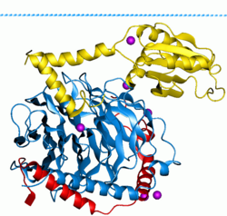
G protein


G proteins, also known as guanine nucleotide-binding proteins, are a family of proteins that act as molecular switches inside cells, and are involved in transmitting signals from a variety of stimuli outside a cell to its interior. Their activity is regulated by factors that control their ability to bind to and hydrolyze guanosine triphosphate (GTP) to guanosine diphosphate (GDP). When they are bound to GTP, they are 'on', and, when they are bound to GDP, they are 'off'. G proteins belong to the larger group of enzymes called GTPases.
There are two classes of G proteins. The first function as
Heterotrimeric G proteins located within the cell are activated by
History
G proteins were discovered in 1980 when
Nobel prizes have been awarded for many aspects of signaling by G proteins and GPCRs. These include
Prominent examples include (in chronological order of awarding):
- The 1947 Carl Cori, Gerty Cori and Bernardo Houssay, for their discovery of how glycogen is broken down to glucose and resynthesized in the body, for use as a store and source of energy. Glycogenolysis is stimulated by numerous hormones and neurotransmitters including adrenaline.
- The 1970 Nobel Prize in Physiology or Medicine to Julius Axelrod, Bernard Katz and Ulf von Euler for their work on the release and reuptake of neurotransmitters.
- The 1971 cyclic AMP.[9]
- The 1988 Gertrude Elion"for their discoveries of important principles for drug treatment" targeting GPCRs.
- The 1992 Nobel Prize in Physiology or Medicine to Edwin G. Krebs and Edmond H. Fischer for describing how reversible phosphorylation works as a switch to activate proteins, and to regulate various cellular processes including glycogenolysis.[11]
- The 1994 Nobel Prize in Physiology or Medicine to Alfred G. Gilman and Martin Rodbell for their discovery of "G-proteins and the role of these proteins in signal transduction in cells".[12]
- The 2000 Nobel Prize in Physiology or Medicine to Eric Kandel, Arvid Carlsson and Paul Greengard, for research on neurotransmitters such as dopamine, which act via GPCRs.
- The 2004 Nobel Prize in Physiology or Medicine to Richard Axel and Linda B. Buck for their work on G protein-coupled olfactory receptors.[13]
- The 2012 Nobel Prize in Chemistry to Brian Kobilka and Robert Lefkowitz for their work on GPCR function.[14]
Function
G proteins are important
Whereas G proteins are activated by
Diversity

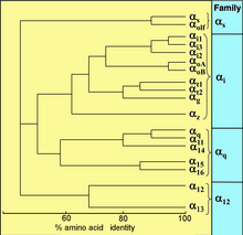
All eukaryotes use G proteins for signaling and have evolved a large diversity of G proteins. For instance, humans encode 18 different Gα proteins, 5 Gβ proteins, and 12 Gγ proteins.[17]
Signaling
G protein can refer to two distinct families of proteins.
Heterotrimeric
Different types of heterotrimeric G proteins share a common mechanism. They are activated in response to a conformational change in the GPCR, exchanging GDP for GTP, and dissociating in order to activate other proteins in a particular signal transduction pathway.[18] The specific mechanisms, however, differ between protein types.
Mechanism

Receptor-activated G proteins are bound to the inner surface of the cell membrane. They consist of the Gα and the tightly associated Gβγ subunits. There are four main families of Gα subunits: Gαs (G stimulatory), Gαi (G inhibitory), Gαq/11, and Gα12/13.[20][21] They behave differently in the recognition of the effector molecule, but share a similar mechanism of activation.
Activation
When a
Termination
The Gα subunit will eventually
Specific mechanisms
Gαs
The cAMP-dependent pathway is used as a signal transduction pathway for many hormones including:
- kidneys (created by the magnocellular neurosecretory cells of the posterior pituitary)
- GHRH – Stimulates the synthesis and release of GH (somatotropic cells of the anterior pituitary)
- GHIH– Inhibits the synthesis and release of GH (somatotropic cells of anterior pituitary)
- CRH – Stimulates the synthesis and release of ACTH (anterior pituitary)
- in the adrenal glands)
- T4(thyroid gland)
- LH – Stimulates follicular maturation and ovulation in women; or testosterone production and spermatogenesis in men
- FSH – Stimulates follicular development in women; or spermatogenesisin men
- blood calcium levels. This is accomplished via the parathyroid hormone 1 receptor (PTH1) in the kidneys and bones, or via the parathyroid hormone 2 receptor(PTH2) in the central nervous system and brain, as well as the bones and kidneys.
- Calcitonin – Decreases blood calcium levels (via the calcitonin receptor in the intestines, bones, kidneys, and brain)
- Glucagon – Stimulates glycogen breakdown in the liver
- hCG – Promotes cellular differentiation, and is potentially involved in apoptosis.[26]
- Epinephrine – released by the adrenal medulla during the fasting state, when body is under metabolic duress. It stimulates glycogenolysis, in addition to the actions of glucagon.
Gαi
Gαq/11
- ADH (Vasopressin/AVP) – Induces the synthesis and release of glucocorticoids (Zona fasciculata of adrenal cortex); Induces vasoconstriction (V1 Cells of Posterior pituitary)
- TRH – Induces the synthesis and release of TSH (Anterior pituitary gland)
- TSH – Induces the synthesis and release of a small amount of T4 (Thyroid Gland)
- Angiotensin II – Induces Aldosterone synthesis and release (zona glomerulosa of adrenal cortex in kidney)
- GnRH – Induces the synthesis and release of FSH and LH (Anterior Pituitary)
Gα12/13
- Gα12/13 are involved in Rho family GTPase signaling (see Rho family of GTPases). This is through the RhoGEF superfamily involving the RhoGEF domain of the proteins' structures). These are involved in control of cell cytoskeleton remodeling, and thus in regulating cell migration.
Gβ, Gγ
- The G protein-coupled inwardly-rectifying potassium channels.
Small GTPases
Small GTPases, also known as small G-proteins, bind GTP and GDP likewise, and are involved in
Lipidation
In order to associate with the inner leaflet[
References
- PMID 10819326.
- PMID 9131251.
- ^ "Seven Transmembrane Receptors: Robert Lefkowitz". 9 September 2012. Retrieved 11 July 2016.
- ^ PMID 21873996.
- PMID 212105.
- PMID 35626696.
- ISBN 0-8053-6624-5.
- S2CID 20136388.
- ^ a b The Nobel Prize in Physiology or Medicine 1994, Illustrated Lecture.
- ^ Press Release: The Nobel Assembly at the Karolinska Institute decided to award the Nobel Prize in Physiology or Medicine for 1994 jointly to Alfred G. Gilman and Martin Rodbell for their discovery of "G-proteins and the role of these proteins in signal transduction in cells". 10 October 1994
- Nobel Assembly at Karolinska Institutet. Retrieved 21 August 2013.
- ^ Press Release
- ^ "Press Release: The 2004 Nobel Prize in Physiology or Medicine". Nobelprize.org. Retrieved 8 November 2012.
- ^ Royal Swedish Academy of Sciences (10 October 2012). "The Nobel Prize in Chemistry 2012 Robert J. Lefkowitz, Brian K. Kobilka". Retrieved 10 October 2012.
- PMID 23519208.
- PMID 23523730.
- ^ PMID 27515397.
- )
- PMID 26123299.
- PMID 27515397.
- ^ "InterPro". www.ebi.ac.uk. Retrieved 25 May 2023.
- PMID 17095603.
- S2CID 23909821.
- PMID 20348012.
- PMID 17854654.
- PMID 20735820.
External links
- GTP-Binding Proteins at the U.S. National Library of Medicine Medical Subject Headings (MeSH)
