
Glomerulus (kidney)
| Glomerulus | |
|---|---|
Metanephric blastema | |
| Location | Nephron of kidney |
| Identifiers | |
| Latin | glomerulus renalis |
| MeSH | D007678 |
| FMA | 15624 |
| Anatomical terminology] | |
The glomerulus (pl.: glomeruli) is a network of small blood vessels (
The glomerulus receives its blood supply from an
The glomerulus and its surrounding
Structure


The glomerulus is a tuft of capillaries located within
Lining

Capillaries of the glomerulus are lined by
The glomerulus has a
The part of the podocyte in contact with the glomerular basement membrane is called a podocyte foot process or pedicle (Fig. 3): there are gaps between the foot processes through which the
Mesangium
The mesangium is a space which is continuous with the smooth muscles of the arterioles. It is outside the capillary lumen but surrounded by capillaries. It is in the middle (meso) between the capillaries (angis). It is contained by the basement membrane, which surrounds both the capillaries and the mesangium.
The mesangium contains mainly:
- pericytesthat participate in the regulation of the filtration rate by contracting or expanding: they contain actin and myosin filaments to accomplish this. Some mesangial cells are in physical contact with capillaries, whereas others are in physical contact with podocytes. There is two-way chemical cross talk among the mesangial cells, the capillaries, and the podocytes to fine-tune the glomerular filtration rate.
- Mesangial matrix, an amorphous basement membrane-like material secreted by the mesangial cells.
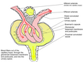
Blood supply

The glomerulus receives its blood supply from an
Blood exits the glomerular capillaries by an
Cortical nephrons near the corticomedullary junction (15% of all nephrons) are called
Filtrate drainage
The filtrate that has passed through the three-layered filtration unit enters Bowman's capsule. From there, it flows into the renal tubule—the nephron—which follows a U-shaped path to the
Function
Filtration

B. Glomerular basement membrane: 1. lamina rara interna 2. lamina densa 3. lamina rara externa
C. Podocytes: 1. enzymatic and structural proteins 2. filtration slit 3. diaphragma
The main function of the glomerulus is to filter
If a substance has passed through the glomerular capillary endothelial cells, glomerular basement membrane, and podocytes, then it enters the lumen of the tubule and is known as glomerular filtrate. Otherwise, it exits the glomerulus through the efferent arteriole and continues circulation as discussed below and as shown on the picture.

Permeability
The structures of the layers determine their
The oncotic pressure on glomerular capillaries is one of the forces that resist filtration. Because large and negatively charged proteins have a low permeability, they cannot filtrate easily to Bowman's capsule. Therefore, the concentration of these proteins tends to increase as the glomerular capillaries filtrate plasma, increasing the oncotic pressure along the glomerular capillary.[6]
Starling equation
The rate of filtration from the glomerulus to Bowman's capsule is determined (as in systemic capillaries) by the Starling equation:[6]

- GFR is the glomerular filtration rate
- Kf is the filtration coefficient—a proportionality constant
- Pgc is the glomerular capillary hydrostatic pressure
- Pbc is the Bowman's capsule hydrostatic pressure
- πgc is the glomerular capillary oncotic pressure
- πbc is the Bowman's capsule oncotic pressure
Blood pressure regulation
The walls of the afferent arteriole contain specialized smooth muscle cells that synthesize renin. These juxtaglomerular cells play a major role in the renin–angiotensin system, which helps regulate blood volume and pressure.
Clinical significance
This section needs expansion. You can help by adding to it. (April 2015) |
Damage to the glomerulus by disease can allow passage through the glomerular filtration barrier of red blood cells, white blood cells, platelets, and blood proteins such as albumin and globulin. Underlying causes for glomerular injury can be inflammatory, toxic or metabolic.
Due to the connection between the glomerulus and the glomerular filtration rate, the glomerular filtration rate is of clinical significance when suspecting a kidney disease, or when following up a case with known kidney disease, or when risking a development of renal damage such as beginning medications with known nephrotoxicity.[10]
History
In 1666, Italian biologist and anatomist Marcello Malpighi first described the glomeruli and demonstrated their continuity with the renal vasculature (281,282). About 175 years later, surgeon and anatomist William Bowman elucidated in detail the capillary architecture of the glomerulus and the continuity between its surrounding capsule and the proximal tubule.[11]
See also
Additional images
-
 Scanning electron microscope image of a glomerulus in a mouse (1000x magnification)
 -
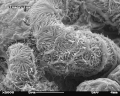 Scanning electron microscope image of a glomerulus in a mouse (5000x magnification)
 -
 Scanning electron microscope image of a glomerulus in a mouse (10,000x magnification)
 -
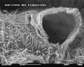
 Looped capillaries of glomerulus between the arterioles


References
- PMID 12506131.
- ^ a b Wheater 2006, p. 304.
- ^ a b Wheater 2006, p. 307.
- ^ a b c Wheater 2006, p. 310.
- PMID 23774818.
- ^ ISBN 978-1437717532.
- ISBN 978-0-7216-0240-0.
- PMID 17410103.
- ^ "Glomerular Diseases: What Is It, Causes, Symptoms & Treatment". Cleveland Clinic. Retrieved 2022-07-27.
- ISBN 978-1-118-34500-9
- ^ "lippicotts histology for pathologesits; satcey e. mills
Sources
- Hall, Arthur C. Guyton, John E. (2005). Textbook of medical physiology (11th ed.). Philadelphia: W.B. Saunders. p. Chapter 26. ISBN 978-0-7216-0240-0.)
{{cite book}}: CS1 maint: multiple names: authors list (link - Deakin, Barbara Young ... [] ; drawings by Philip J.; et al. (2006). Wheater's Functional Histology: a text and colour atlas (5th ed.). [Edinburgh?]: Churchill Livingstone/Elsevier. p. Chapter 16. ISBN 978-0-443068508.)
{{cite book}}: CS1 maint: multiple names: authors list (link