
Glossopharyngeal nerve
| Glossopharyngeal nerve | |
|---|---|
Oropharynx, Eustachian tube, middle ear, posterior third of tongue, carotid sinus, carotid body Special sensory: Taste to posterior third of tongue | |
| Identifiers | |
| Latin | nervus glossopharyngeus |
| MeSH | D005930 |
| NeuroNames | 701, 793 |
| NeuroLex ID | birnlex_1274 |
| TA98 | A14.2.01.135 |
| TA2 | 6320 |
| FMA | 50870 |
| Anatomical terms of neuroanatomy] | |
| Cranial nerves |
|---|
|
The glossopharyngeal nerve (
Structure
From the anterior portion of the
In its passage through the foramen (with X and XI), the glossopharyngeal nerve passes between the
.Branches
- tympanic nerve
- stylopharyngeal nerve
- tonsillar nerve
- carotid sinus nerve
- Branches to the posterior third of tongue
- lingual branches
- A communicating branch to the vagus nerve
Note: The glossopharyngeal nerve contributes in the formation of the pharyngeal plexus along with the vagus nerve.
The glossopharyngeal nerve has five distinct general functions:
- Branchial motor (special visceral efferent) – supplies the stylopharyngeus muscle.
- Visceral motor (parasympathetic innervation of the parotid glandvia the otic ganglion
- Visceral sensory (general visceral afferent) – carries visceral sensory information from the carotid sinus and carotid body.
- General sensory (.
- Visceral afferent (circumvallate papillae.
The glossopharyngeal nerve as noted above is a mixed nerve consisting of both sensory and motor nerve fibers. The sensory fibers' origin include the pharynx, middle ear, posterior one-third of the tongue (including taste buds); and the carotid body and sinus. These fibers terminate at the
Overview of branchial motor component
The
Origin and central course
The branchial motor component originates from the nucleus ambiguus in the reticular formation of the rostral medulla. Fibers leaving the nucleus ambiguus travel anteriorly and laterally to exit the medulla, along with the other components of CN IX, between the olive and the inferior cerebellar peduncle.
Intracranial course
Upon emerging from the lateral aspect of the medulla the branchial motor component joins the other components of CN IX to exit the skull via the jugular foramen. The glossopharyngeal fibers travel just anterior to the cranial nerves X and XI, which also exit the skull via the jugular foramen.
Extra-cranial course and final innervation
Upon exiting the skull the branchial motor fibers descend deep to the temporal styloid process and wrap around the posterior border of the stylopharyngeus muscle before innervating it.
Voluntary control of the stylopharyngeus muscle
Signals for the voluntary movement of
Overview of visceral motor component
Parasympathetic component of the glossopharyngeal nerve that innervates the ipsilateral parotid gland.
Origin and central course
The
Intracranial course
Upon emerging from the lateral aspect of the medulla, the
Extra-cranial course and final innervations
Upon exiting the skull, the lesser petrosal nerve synapses in the
Hypothalamic Influence
Fibers from the
Overview of visceral sensory component
This component of CN IX innervates the baroreceptors of the carotid sinus and chemoreceptors of the carotid body.
- Peripheral and intracranial course.
- Sensory fibers arise from the CN IX at the inferior glossopharyngeal ganglion. The cell bodies of these neurons reside in the inferior glossopharyngeal ganglion. The central processes of these neurons enter the skull via the jugular foramen.
- Central course – visceral sensory component
- Once inside the skull, the visceral sensory fibers enter the lateral medulla between the olive and the solitary nucleus. From the solitary nucleus, connections are made with several areas in the reticular formation and hypothalamusto mediate cardiovascular and respiratory reflex responses to changes in blood pressure, and serum concentrations of CO2 and O2.
Clinical correlation The visceral sensory fibers of CN IX mediate the afferent limb of the pharyngeal reflex in which touching the back of the pharynx stimulates the patient to gag (i.e., the gag reflex). The efferent signal to the musculature of the pharynx is carried by the branchial motor fibers of the vagus nerve. [5]
Overview of somatic sensory component
This component of CN IX carries general sensory information (pain, temperature, and touch) from the skin of the external ear, internal surface of the tympanic membrane, the walls of the upper pharynx, and the posterior one-third of the tongue, anterior surface of the epiglottis, vallecula.
- Peripheral course
- Sensory fibers from the skin of the external ear initially travel with the auricular branch of CN X, while those from the middle ear travel in the tympanic nerve as discussed above (CN IX visceral motor section). General sensory information from the upper pharynx and posterior one-third of the tongue travel via the pharyngeal branches of CN IX. These peripheral processes have their cell body in either the superior or inferior glossopharyngeal ganglion.
- Central course
- The central processes of the general sensory neurons exit the glossopharyngeal ganglia and pass through the jugular foramen to enter the brainstem at the level of the medulla. Upon entering the medulla these fibers descend in the spinal trigeminal tract and synapse in the caudal spinal nucleus of the trigeminal.
Overview of special sensory component
The special sensory component of CN IX provides taste sensation from the posterior one-third of the tongue.
- Peripheral course
- Special sensory fibers from the posterior one-third of the tongue travel via the pharyngeal branches of CN IX to the inferior glossopharyngeal ganglion where their cell bodies reside.
- Central course – special sensory component
- The central processes of these neurons exit the inferior ganglion and pass through the jugular foramen to enter the brainstem at the level of the rostral medulla between the olive and inferior cerebellar peduncle. Upon entering the medulla, these fibers ascend in the tractus solitarius and synapse in the gustatory part of nucleus solitarius. Taste fibers from CN VII and X also ascend and synapse here. Ascending secondary neurons originating in nucleus solitarius project bilaterally to the ventral posteromedial (VPM) nuclei of the thalamus via the central tegmental tract. Tertiary neurons from the thalamus project via the posterior limb of the internal capsule to the inferior one-third of the primary sensory cortex (the gustatory cortex of the parietal lobe).
Associated brainstem nuclei
- Solitary nucleus: taste from the posterior 1/3 of the tongue and information from carotid sinus baroreceptors and carotid body chemoreceptors
- Spinal nucleus of the trigeminal nerve: Somatic sensory fibers from the internal surface of the tympanic membrane, middle ear, upper part of the pharynx, soft palate and posterior 1/3 of the tongue
- stylopharyngeusmuscle
- Inferior salivatory nucleus: preganglionic parasympatheticneurons to the otic ganglion and then to the parotid gland
Functions
- It receives general somatic sensory fibers (tonsils, the pharynx, the middle earand the posterior 1/3 of the tongue.
- It receives special visceral sensory fibers (taste) from the posterior 1/3 of the tongue.
- It receives visceral sensory fibers from the carotid bodies, carotid sinus.[6]
- It supplies
- It supplies motor fibers to stylopharyngeus muscle, the only motor component of this cranial nerve.[2]
- It contributes to the pharyngeal plexus.
Clinical significance
Damage
Damage to the glossopharyngeal nerve can result in loss of taste sensation to the posterior one third of the tongue, and impaired swallowing.
Examination
The clinical tests used to determine if the glossopharyngeal nerve has been damaged include testing the
, and evaluating for speech impediments. The clinician may also test the posterior one-third of the tongue with bitter and sour substances to evaluate for impairment of taste.The integrity of the glossopharyngeal nerve may be evaluated by testing the patient's general sensation and that of taste on the posterior third of the tongue. The gag reflex can also be used to evaluate the glossphyaryngeal nerve.
Additional images
-
 Inferior view of the human brain, with the cranial nerves labelled.
 -
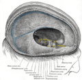 Dura mater and its processes exposed by removing part of the right half of the skull, and the brain.
 -
 Hind- and mid-brains; postero-lateral view.(
 -
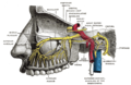 Alveolar branches of superior maxillary nerve and sphenopalatine ganglion.
 -
 Upper part of medulla spinalis and hind- and mid-brains; posterior aspect, exposed in situ.
 -
 Hypoglossal nerve, cervical plexus, and their branches.


References
- ^ "Glossopharyngeal | Definition of Glossopharyngeal by Oxford Dictionary on Lexico.com also meaning of Glossopharyngeal". Lexico Dictionaries | English. Archived from the original on March 12, 2021.
- ^ ISBN 978-0-12-226870-0, retrieved 2020-12-31
- ISBN 978-0-323-37704-1, retrieved 2020-12-31
- ISBN 978-0-7020-3100-7, retrieved 2020-12-31
- .
- )
Saladin, Anatomy and Physiology: The Unity of Form and Function, 6th edition
External links
- hier-698 at NeuroNames
- MedEd at Loyola GrossAnatomy/h_n/cn/cn1/cn9.htm
- MedlinePlus Image 9350
- cranialnerves at The Anatomy Lesson by Wesley Norman (Georgetown University) (IX)